 Movie
Movie Controller


+ Open data

- Basic information
| Entry | Database: PDB / ID: 9x6s | ||||||||||||
|---|---|---|---|---|---|---|---|---|---|---|---|---|---|
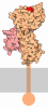
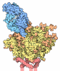
| Title | Structure of Influenza Hemagglutinin (A/Puerto Rico/8/1934) | ||||||||||||
 Components Components | Hemagglutinin | ||||||||||||
 Keywords Keywords | VIRAL PROTEIN / Hemagglutinin / Glycoprotein.Prefusion / Ectodomain | ||||||||||||
| Function / homology |  Function and homology information Function and homology informationTransport of HA trimer, NA tetramer and M2 tetramer from the endoplasmic reticulum to the Golgi Apparatus / Assembly of Viral Components at the Budding Site / Influenza Infection / Fusion of the Influenza Virion to the Host Cell Endosome / Release / Budding / Packaging of Eight RNA Segments / Uncoating of the Influenza Virion / Entry of Influenza Virion into Host Cell via Endocytosis / Viral mRNA Translation ...Transport of HA trimer, NA tetramer and M2 tetramer from the endoplasmic reticulum to the Golgi Apparatus / Assembly of Viral Components at the Budding Site / Influenza Infection / Fusion of the Influenza Virion to the Host Cell Endosome / Release / Budding / Packaging of Eight RNA Segments / Uncoating of the Influenza Virion / Entry of Influenza Virion into Host Cell via Endocytosis / Viral mRNA Translation / viral budding from plasma membrane / Immunoregulatory interactions between a Lymphoid and a non-Lymphoid cell / clathrin-dependent endocytosis of virus by host cell / host cell surface receptor binding / fusion of virus membrane with host plasma membrane / fusion of virus membrane with host endosome membrane / viral envelope / virion attachment to host cell / host cell plasma membrane / virion membrane / extracellular region / plasma membrane Similarity search - Function | ||||||||||||
| Biological species |   Influenza A virus Influenza A virus | ||||||||||||
| Method | ELECTRON MICROSCOPY / subtomogram averaging / cryo EM / Resolution: 3.6 Å | ||||||||||||
 Authors Authors | Chen, Y. / Li, S. | ||||||||||||
| Funding support |  China, 3items China, 3items
| ||||||||||||
 Citation Citation | Journal: Nat Commun / Year: 2026 Title: FlyTomo: a streamlined software for on-the-fly cryo-ET data processing and diagnosis. Authors: Zheyuan Zhang / Cheng Peng / Weiping Zhang / Jiaming Liang / Kexin Liu / Yong Chen / Junxia Zhang / Rui Liang / Yutong Song / Sai Li / Abstract: Cryo-ET combined with subtomogram averaging (STA) enables the structural elucidation of macromolecular assemblies in native environments. However, their widespread adoption has been limited by the ...Cryo-ET combined with subtomogram averaging (STA) enables the structural elucidation of macromolecular assemblies in native environments. However, their widespread adoption has been limited by the labor-intensive, expertise-dependent data processing workflow. Here we present FlyTomo, a software that streamlines data processing from frame alignment to STA with high-throughput for authentic cryo-ET scenarios. During data acquisition, FlyTomo performs real-time diagnosis, enabling prompt feedback on sample quality, microscope performance and structural features. After acquisition, it aggregates diagnostic metrics into an overview, guiding users through data review and refinement. FlyTomo also curates raw and processed data into directories to simplify data management and archiving. We validate FlyTomo across a diverse set of authentic cryo-ET samples, including purified enveloped viruses and cryo-lamellae, on multiple microscopes and cameras, achieving structures at resolutions of 3.4 to 7.3 Å. Collectively, by integrating accuracy, scalability and usability, FlyTomo reduces the technical barrier for in situ structural biology using cryo-ET. | ||||||||||||
| History |
|
- Structure visualization
| Structure viewer | Molecule: MolmilJmol/JSmol |
|---|
- Downloads & links
-Download
| PDBx/mmCIF format | 9x6s.cif.gz | 269.2 KB | Display | PDBx/mmCIF format |
|---|---|---|---|---|
| PDB format | pdb9x6s.ent.gz | 219.9 KB | Display | PDB format |
| PDBx/mmJSON format | 9x6s.json.gz | Tree view | PDBx/mmJSON format | |
| Others |  Other downloads Other downloads |
-Validation report
| Arichive directory | https://data.pdbj.org/pub/pdb/validation_reports/x6/9x6sftp://data.pdbj.org/pub/pdb/validation_reports/x6/9x6s | HTTPS FTP |
|---|
-Related structure data
| Related structure data |  66624MC  9x6zC M: map data used to model this data C: citing same article ( |
|---|---|
| Similar structure data |


-Links
 PDBj
PDBj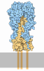


- Assembly
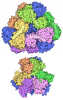
| Deposited unit | 
|
|---|---|
| 1 |
|
-Components
| #1: Protein | Mass: 63422.621 Da / Num. of mol.: 3 / Source method: isolated from a natural source Source: (natural) Influenza A virus (A/Puerto Rico/8/1934(H1N1))Strain: A/Puerto Rico/8/1934(H1N1) / References: UniProt: P03452 #2: Sugar | ChemComp-NAG /   Type: D-saccharide, beta linking / Mass: 221.208 Da / Num. of mol.: 15 / Source method: obtained synthetically / Formula: C8H15NO6 / Feature type: SUBJECT OF INVESTIGATION Type: D-saccharide, beta linking / Mass: 221.208 Da / Num. of mol.: 15 / Source method: obtained synthetically / Formula: C8H15NO6 / Feature type: SUBJECT OF INVESTIGATIONHas ligand of interest | Y | Has protein modification | Y | |
|---|
-Experimental details
-Experiment
| Experiment | Method: ELECTRON MICROSCOPY |
|---|---|
| EM experiment | Aggregation state: PARTICLE / 3D reconstruction method: subtomogram averaging |
- Sample preparation
| Component | Name: Hemagglutinin protein on virial surface / Type: COMPLEX / Entity ID: #1 / Source: NATURAL |
|---|---|
| Source (natural) | Organism: Influenza A virus / Strain: A/Puerto Rico/8/1934(H1N1) |
| Buffer solution | pH: 7.4 |
| Specimen | Embedding applied: NO / Shadowing applied: NO / Staining applied: NO / Vitrification applied: YES |
| Vitrification | Cryogen name: ETHANE |
- Electron microscopy imaging
| Experimental equipment |  Model: Titan Krios / Image courtesy: FEI Company |
|---|---|
| Microscopy | Model: TFS KRIOS |
| Electron gun | Electron source:  FIELD EMISSION GUN / Accelerating voltage: 300 kV / Illumination mode: FLOOD BEAM FIELD EMISSION GUN / Accelerating voltage: 300 kV / Illumination mode: FLOOD BEAM |
| Electron lens | Mode: BRIGHT FIELD / Nominal defocus max: 4000 nm / Nominal defocus min: 2000 nm |
| Image recording | Electron dose: 3.2 e/Å2 / Avg electron dose per subtomogram: 131.2 e/Å2 / Film or detector model: FEI FALCON IV (4k x 4k) |
- Processing
| EM software |
| ||||||||||||
|---|---|---|---|---|---|---|---|---|---|---|---|---|---|
| CTF correction | Type: PHASE FLIPPING ONLY | ||||||||||||
| Symmetry | Point symmetry: C3 (3 fold cyclic) | ||||||||||||
| 3D reconstruction | Resolution: 3.6 Å / Resolution method: FSC 0.143 CUT-OFF / Num. of particles: 97201 / Symmetry type: POINT | ||||||||||||
| EM volume selection | Num. of tomograms: 65 / Num. of volumes extracted: 260571 |
