 Movie
Movie Controller


+ Open data

- Basic information
| Entry | Database: PDB / ID: 9x4e | |||||||||
|---|---|---|---|---|---|---|---|---|---|---|
| Title | Structure of the old Killifish Proteasome | |||||||||
 Components Components | (Proteasome subunit ...) x 14 | |||||||||
 Keywords Keywords | HYDROLASE / Proteasome / Nothobranchius furzeri / aging / skeletal muscle | |||||||||
| Function / homology |  Function and homology information Function and homology informationproteasome core complex / proteasome core complex, beta-subunit complex / threonine-type endopeptidase activity / proteasome core complex, alpha-subunit complex / proteasomal protein catabolic process / protein catabolic process / ubiquitin-dependent protein catabolic process / proteasome-mediated ubiquitin-dependent protein catabolic process / nucleus / cytosol / cytoplasm Similarity search - Function | |||||||||
| Biological species |  Nothobranchius furzeri (turquoise killifish) Nothobranchius furzeri (turquoise killifish) | |||||||||
| Method | ELECTRON MICROSCOPY / single particle reconstruction / cryo EM / Resolution: 3.26 Å | |||||||||
 Authors Authors | Kim, J. / Song, J.J. / Kim, Y. | |||||||||
| Funding support |  Korea, Republic Of, 2items Korea, Republic Of, 2items
| |||||||||
 Citation Citation | Journal: To Be Published Title: Ribosome remodels in age-dependent manner Authors: Kim, J. / Hyeon, D.Y. / Lee, S. / Ryu, B.H. / Gwak, N. / Choi, Y. / Vu, H.M. / Huh, S. / Kim, S. / Myung, K. / Dieterich, C. / Valenzano, D.R. / Kim, M.S. / Hwang, D. / Song, J.J. / Kim, Y. | |||||||||
| History |
|
- Structure visualization
| Structure viewer | Molecule: MolmilJmol/JSmol |
|---|
- Downloads & links
-Download
| PDBx/mmCIF format | 9x4e.cif.gz | 1.2 MB | Display | PDBx/mmCIF format |
|---|---|---|---|---|
| PDB format | pdb9x4e.ent.gz | 957.3 KB | Display | PDB format |
| PDBx/mmJSON format | 9x4e.json.gz | Tree view | PDBx/mmJSON format | |
| Others |  Other downloads Other downloads |
-Validation report
| Arichive directory | https://data.pdbj.org/pub/pdb/validation_reports/x4/9x4eftp://data.pdbj.org/pub/pdb/validation_reports/x4/9x4e | HTTPS FTP |
|---|
-Related structure data
| Related structure data |  66543MC  9x4dC  9x4iC M: map data used to model this data C: citing same article ( |
|---|---|
| Similar structure data |


-Links
 PDBj
PDBj

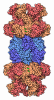
- Assembly
| Deposited unit | 
|
|---|---|
| 1 |
|
-Components
-Proteasome subunit ... , 14 types, 28 molecules AOBPCQDRESFTGUHXIWJVKZLYMaNb
| #1: Protein | Mass: 25900.441 Da / Num. of mol.: 2 / Source method: isolated from a natural source Source: (natural) Nothobranchius furzeri (turquoise killifish)Tissue: skeletal muscle / References: UniProt: A0A1A7ZN72 #2: Protein | Mass: 29405.709 Da / Num. of mol.: 2 / Source method: isolated from a natural source Source: (natural) Nothobranchius furzeri (turquoise killifish)Tissue: skeletal muscle / References: UniProt: A0A1A8B0E3 #3: Protein | Mass: 28143.230 Da / Num. of mol.: 2 / Source method: isolated from a natural source Source: (natural) Nothobranchius furzeri (turquoise killifish)Tissue: skeletal muscle / References: UniProt: A0A1A8V7S3 #4: Protein | Mass: 26445.910 Da / Num. of mol.: 2 / Source method: isolated from a natural source Source: (natural) Nothobranchius furzeri (turquoise killifish)Tissue: skeletal muscle / References: UniProt: A0A1A8V3Y9 #5: Protein | Mass: 29380.420 Da / Num. of mol.: 2 / Source method: isolated from a natural source Source: (natural) Nothobranchius furzeri (turquoise killifish)Tissue: skeletal muscle / References: UniProt: A0A1A8UYX9 #6: Protein | Mass: 28428.021 Da / Num. of mol.: 2 / Source method: isolated from a natural source Source: (natural) Nothobranchius furzeri (turquoise killifish)Tissue: skeletal muscle / References: UniProt: A0A1A7ZU17 #7: Protein | Mass: 27471.342 Da / Num. of mol.: 2 / Source method: isolated from a natural source Source: (natural) Nothobranchius furzeri (turquoise killifish)Tissue: skeletal muscle / References: UniProt: A0A1A8U241 #8: Protein | Mass: 25022.373 Da / Num. of mol.: 2 / Source method: isolated from a natural source Source: (natural) Nothobranchius furzeri (turquoise killifish)Tissue: skeletal muscle / References: UniProt: A0A1A8A9L4 #9: Protein | Mass: 23050.857 Da / Num. of mol.: 2 / Source method: isolated from a natural source Source: (natural) Nothobranchius furzeri (turquoise killifish)Tissue: skeletal muscle / References: UniProt: A0A1A8ANK7 #10: Protein | Mass: 29726.891 Da / Num. of mol.: 2 / Source method: isolated from a natural source Source: (natural) Nothobranchius furzeri (turquoise killifish)Tissue: skeletal muscle / References: UniProt: A0A1A8TWU4 #11: Protein | Mass: 32160.512 Da / Num. of mol.: 2 / Source method: isolated from a natural source / Details: Sequence refers to XP_054589013.1 Source: (natural) Nothobranchius furzeri (turquoise killifish)Tissue: skeletal muscle #12: Protein | Mass: 23931.572 Da / Num. of mol.: 2 / Source method: isolated from a natural source Source: (natural) Nothobranchius furzeri (turquoise killifish)Tissue: skeletal muscle / References: UniProt: A0A1A7ZAT0 #13: Protein | Mass: 28307.088 Da / Num. of mol.: 2 / Source method: isolated from a natural source Source: (natural) Nothobranchius furzeri (turquoise killifish)Tissue: skeletal muscle / References: UniProt: A0A1A8AVF5 #14: Protein | Mass: 26073.697 Da / Num. of mol.: 2 / Source method: isolated from a natural source Source: (natural) Nothobranchius furzeri (turquoise killifish)Tissue: skeletal muscle / References: UniProt: A0A1A8UKL0 |
|---|
-Details
| Has protein modification | N |
|---|
-Experimental details
-Experiment
| Experiment | Method: ELECTRON MICROSCOPY |
|---|---|
| EM experiment | Aggregation state: PARTICLE / 3D reconstruction method: single particle reconstruction |
- Sample preparation
| Component | Name: Structure of the old Killifish Proteasome / Type: COMPLEX / Entity ID: all / Source: NATURAL | ||||||||||||||||||||
|---|---|---|---|---|---|---|---|---|---|---|---|---|---|---|---|---|---|---|---|---|---|
| Source (natural) | Organism: Nothobranchius furzeri (turquoise killifish) / Tissue: skeletal muscle | ||||||||||||||||||||
| Buffer solution | pH: 7.5 | ||||||||||||||||||||
| Buffer component |
| ||||||||||||||||||||
| Specimen | Embedding applied: NO / Shadowing applied: NO / Staining applied: NO / Vitrification applied: YES | ||||||||||||||||||||
| Specimen support | Grid material: COPPER / Grid mesh size: 300 divisions/in. / Grid type: Quantifoil R2/2 | ||||||||||||||||||||
| Vitrification | Cryogen name: ETHANE |
- Electron microscopy imaging
| Experimental equipment |  Model: Titan Krios / Image courtesy: FEI Company |
|---|---|
| Microscopy | Model: TFS KRIOS |
| Electron gun | Electron source:  FIELD EMISSION GUN / Accelerating voltage: 300 kV / Illumination mode: FLOOD BEAM FIELD EMISSION GUN / Accelerating voltage: 300 kV / Illumination mode: FLOOD BEAM |
| Electron lens | Mode: BRIGHT FIELD / Nominal defocus max: 1000 nm / Nominal defocus min: 300 nm / Cs: 2.7 mm / C2 aperture diameter: 70 µm |
| Specimen holder | Cryogen: NITROGEN |
| Image recording | Average exposure time: 5.87 sec. / Electron dose: 60.23 e/Å2 / Film or detector model: GATAN K3 BIOQUANTUM (6k x 4k) / Num. of real images: 9136 |
| EM imaging optics | Energyfilter name: GIF Bioquantum / Details: BioQuantum K3 Imaging filter / Energyfilter slit width: 20 eV |
- Processing
| EM software |
| |||||||||||||||||||||||||||||||||||
|---|---|---|---|---|---|---|---|---|---|---|---|---|---|---|---|---|---|---|---|---|---|---|---|---|---|---|---|---|---|---|---|---|---|---|---|---|
| CTF correction | Type: PHASE FLIPPING AND AMPLITUDE CORRECTION | |||||||||||||||||||||||||||||||||||
| Particle selection | Num. of particles selected: 2953254 | |||||||||||||||||||||||||||||||||||
| Symmetry | Point symmetry: C1 (asymmetric) | |||||||||||||||||||||||||||||||||||
| 3D reconstruction | Resolution: 3.26 Å / Resolution method: FSC 0.143 CUT-OFF / Num. of particles: 121745 / Symmetry type: POINT | |||||||||||||||||||||||||||||||||||
| Refinement | Cross valid method: NONE |
