Movie
Movie Controller

[English] 日本語
 Yorodumi
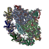
- PDB-9wls: PSI complex of A.thaliana isolated using DOC based Clear-Native-P... -
+ Open data

- Basic information
| Entry | Database: PDB / ID: 9wls | ||||||||||||||||||||||||||||||
|---|---|---|---|---|---|---|---|---|---|---|---|---|---|---|---|---|---|---|---|---|---|---|---|---|---|---|---|---|---|---|---|
| Title | PSI complex of A.thaliana isolated using DOC based Clear-Native-PAGE method | ||||||||||||||||||||||||||||||
 Components Components |
| ||||||||||||||||||||||||||||||
 Keywords Keywords | PHOTOSYNTHESIS / Photosystem I complex / membrane protein | ||||||||||||||||||||||||||||||
| Function / homology |  Function and homology information Function and homology informationphotosynthetic NADP+ reduction / photosystem I stabilization / chloroplast photosystem I / chloroplast stromal thylakoid / response to low light intensity stimulus / plastoglobule / chloroplast membrane / response to high light intensity / chloroplast thylakoid / photosynthesis, light harvesting in photosystem I ...photosynthetic NADP+ reduction / photosystem I stabilization / chloroplast photosystem I / chloroplast stromal thylakoid / response to low light intensity stimulus / plastoglobule / chloroplast membrane / response to high light intensity / chloroplast thylakoid / photosynthesis, light harvesting in photosystem I / chloroplast thylakoid lumen / thylakoid / chloroplast envelope / photosystem I reaction center / photosystem I / photosynthetic electron transport in photosystem I / photosystem I / plastid / chlorophyll binding / chloroplast thylakoid membrane / response to light stimulus / photosynthesis / response to cold / chloroplast / 4 iron, 4 sulfur cluster binding / calmodulin binding / electron transfer activity / oxidoreductase activity / protein stabilization / protein domain specific binding / mRNA binding / magnesium ion binding / extracellular region / metal ion binding / nucleus / plasma membrane / cytosol Similarity search - Function | ||||||||||||||||||||||||||||||
| Biological species |  | ||||||||||||||||||||||||||||||
| Method | ELECTRON MICROSCOPY / single particle reconstruction / cryo EM / Resolution: 2.18 Å | ||||||||||||||||||||||||||||||
 Authors Authors | Kawamoto, A. / Seki, S. / Kurisu, G. | ||||||||||||||||||||||||||||||
| Funding support |  Japan, 2items Japan, 2items
| ||||||||||||||||||||||||||||||
 Citation Citation | Journal: To Be Published Title: A novel method for cryo-EM protein sample preparation based on Clear-Native-PAGE Authors: Yang, Z. / Kameo, S. / Seki, S. / Kurisu, G. / Tanaka, R. / Kawamoto, A. / Takabayashi, A. #1: Journal: Acta Crystallogr D Struct Biol / Year: 2019 Title: Macromolecular structure determination using X-rays, neutrons and electrons: recent developments in Phenix. Authors: Dorothee Liebschner / Pavel V Afonine / Matthew L Baker / Gábor Bunkóczi / Vincent B Chen / Tristan I Croll / Bradley Hintze / Li Wei Hung / Swati Jain / Airlie J McCoy / Nigel W Moriarty ...Authors: Dorothee Liebschner / Pavel V Afonine / Matthew L Baker / Gábor Bunkóczi / Vincent B Chen / Tristan I Croll / Bradley Hintze / Li Wei Hung / Swati Jain / Airlie J McCoy / Nigel W Moriarty / Robert D Oeffner / Billy K Poon / Michael G Prisant / Randy J Read / Jane S Richardson / David C Richardson / Massimo D Sammito / Oleg V Sobolev / Duncan H Stockwell / Thomas C Terwilliger / Alexandre G Urzhumtsev / Lizbeth L Videau / Christopher J Williams / Paul D Adams /    Abstract: Diffraction (X-ray, neutron and electron) and electron cryo-microscopy are powerful methods to determine three-dimensional macromolecular structures, which are required to understand biological ...Diffraction (X-ray, neutron and electron) and electron cryo-microscopy are powerful methods to determine three-dimensional macromolecular structures, which are required to understand biological processes and to develop new therapeutics against diseases. The overall structure-solution workflow is similar for these techniques, but nuances exist because the properties of the reduced experimental data are different. Software tools for structure determination should therefore be tailored for each method. Phenix is a comprehensive software package for macromolecular structure determination that handles data from any of these techniques. Tasks performed with Phenix include data-quality assessment, map improvement, model building, the validation/rebuilding/refinement cycle and deposition. Each tool caters to the type of experimental data. The design of Phenix emphasizes the automation of procedures, where possible, to minimize repetitive and time-consuming manual tasks, while default parameters are chosen to encourage best practice. A graphical user interface provides access to many command-line features of Phenix and streamlines the transition between programs, project tracking and re-running of previous tasks. | ||||||||||||||||||||||||||||||
| History |
|
- Structure visualization
| Structure viewer | Molecule: MolmilJmol/JSmol |
|---|
- Downloads & links
-Download
| PDBx/mmCIF format | 9wls.cif.gz | 1.2 MB | Display | PDBx/mmCIF format |
|---|---|---|---|---|
| PDB format | pdb9wls.ent.gz | Display | PDB format | |
| PDBx/mmJSON format | 9wls.json.gz | Tree view | PDBx/mmJSON format | |
| Others |  Other downloads Other downloads |
-Validation report
| Arichive directory | https://data.pdbj.org/pub/pdb/validation_reports/wl/9wlsftp://data.pdbj.org/pub/pdb/validation_reports/wl/9wls | HTTPS FTP |
|---|
-Related structure data
| Related structure data |  66073MC M: map data used to model this data C: citing same article ( |
|---|---|
| Similar structure data |
-Links
 PDBj
PDBj


- Assembly
| Deposited unit | 
|
|---|---|
| 1 |
|
-Components
-Chlorophyll a-b binding protein ... , 2 types, 2 molecules 14
| #1: Protein | Mass: 26021.895 Da / Num. of mol.: 1 / Source method: isolated from a natural source / Source: (natural) |
|---|---|
| #4: Protein | Mass: 27760.461 Da / Num. of mol.: 1 / Source method: isolated from a natural source / Source: (natural) |
-Photosystem I chlorophyll a/b-binding protein ... , 2 types, 2 molecules 23
| #2: Protein | Mass: 27782.814 Da / Num. of mol.: 1 / Source method: isolated from a natural source / Source: (natural) |
|---|---|
| #3: Protein | Mass: 29206.311 Da / Num. of mol.: 1 / Source method: isolated from a natural source / Source: (natural) |
-Photosystem I P700 chlorophyll a apoprotein ... , 2 types, 2 molecules AB
| #5: Protein | Mass: 83315.367 Da / Num. of mol.: 1 / Source method: isolated from a natural source / Source: (natural) |
|---|---|
| #6: Protein | Mass: 82555.883 Da / Num. of mol.: 1 / Source method: isolated from a natural source / Source: (natural) |
-Photosystem I reaction center subunit ... , 10 types, 10 molecules DEFGHIJKLN
| #8: Protein | Mass: 22336.598 Da / Num. of mol.: 1 / Source method: isolated from a natural source / Source: (natural) |
|---|---|
| #9: Protein | Mass: 14984.955 Da / Num. of mol.: 1 / Source method: isolated from a natural source / Source: (natural) |
| #10: Protein | Mass: 24203.125 Da / Num. of mol.: 1 / Source method: isolated from a natural source / Source: (natural) |
| #11: Protein | Mass: 17103.271 Da / Num. of mol.: 1 / Source method: isolated from a natural source / Source: (natural) |
| #12: Protein | Mass: 15291.522 Da / Num. of mol.: 1 / Source method: isolated from a natural source / Source: (natural) |
| #13: Protein/peptide | Mass: 4137.024 Da / Num. of mol.: 1 / Source method: isolated from a natural source / Source: (natural) |
| #14: Protein/peptide | Mass: 5011.897 Da / Num. of mol.: 1 / Source method: isolated from a natural source / Source: (natural) |
| #15: Protein | Mass: 13219.431 Da / Num. of mol.: 1 / Source method: isolated from a natural source / Source: (natural) |
| #16: Protein | Mass: 23070.557 Da / Num. of mol.: 1 / Source method: isolated from a natural source / Source: (natural) |
| #17: Protein | Mass: 18451.064 Da / Num. of mol.: 1 / Source method: isolated from a natural source / Source: (natural) |
-Protein / Sugars , 2 types, 2 molecules C
| #29: Sugar | ChemComp-DGD /  Type: saccharide / Mass: 949.299 Da / Num. of mol.: 1 / Source method: obtained synthetically / Formula: C51H96O15 / Feature type: SUBJECT OF INVESTIGATION Type: saccharide / Mass: 949.299 Da / Num. of mol.: 1 / Source method: obtained synthetically / Formula: C51H96O15 / Feature type: SUBJECT OF INVESTIGATION |
|---|---|
| #7: Protein | Mass: 9049.509 Da / Num. of mol.: 1 / Source method: isolated from a natural source / Source: (natural) |
-Non-polymers , 12 types, 685 molecules 

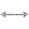


| #18: Chemical | ChemComp-CHL /  Mass: 907.472 Da / Num. of mol.: 9 / Source method: obtained synthetically / Formula: C55H70MgN4O6 / Feature type: SUBJECT OF INVESTIGATION Mass: 907.472 Da / Num. of mol.: 9 / Source method: obtained synthetically / Formula: C55H70MgN4O6 / Feature type: SUBJECT OF INVESTIGATION#19: Chemical | ChemComp-CLA /  Mass: 893.489 Da / Num. of mol.: 117 / Source method: obtained synthetically / Formula: C55H72MgN4O5 / Feature type: SUBJECT OF INVESTIGATION Mass: 893.489 Da / Num. of mol.: 117 / Source method: obtained synthetically / Formula: C55H72MgN4O5 / Feature type: SUBJECT OF INVESTIGATION#20: Chemical | ChemComp-UNL / Num. of mol.: 29 / Source method: obtained synthetically / Feature type: SUBJECT OF INVESTIGATION #21: Chemical |  Mass: 600.870 Da / Num. of mol.: 3 / Source method: obtained synthetically / Formula: C40H56O4 / Feature type: SUBJECT OF INVESTIGATION Mass: 600.870 Da / Num. of mol.: 3 / Source method: obtained synthetically / Formula: C40H56O4 / Feature type: SUBJECT OF INVESTIGATION#22: Chemical | ChemComp-A1LXP / Mass: 568.871 Da / Num. of mol.: 8 / Source method: obtained synthetically / Formula: C40H56O2 / Feature type: SUBJECT OF INVESTIGATION #23: Chemical | ChemComp-LHG /  Mass: 722.970 Da / Num. of mol.: 4 / Source method: obtained synthetically / Formula: C38H75O10P / Feature type: SUBJECT OF INVESTIGATION / Comment: phospholipid*YM Mass: 722.970 Da / Num. of mol.: 4 / Source method: obtained synthetically / Formula: C38H75O10P / Feature type: SUBJECT OF INVESTIGATION / Comment: phospholipid*YM#24: Chemical | ChemComp-BCR /  Mass: 536.873 Da / Num. of mol.: 23 / Source method: obtained synthetically / Formula: C40H56 / Feature type: SUBJECT OF INVESTIGATION Mass: 536.873 Da / Num. of mol.: 23 / Source method: obtained synthetically / Formula: C40H56 / Feature type: SUBJECT OF INVESTIGATION#25: Chemical | ChemComp-LMG /  Mass: 787.158 Da / Num. of mol.: 6 / Source method: obtained synthetically / Formula: C45H86O10 / Feature type: SUBJECT OF INVESTIGATION Mass: 787.158 Da / Num. of mol.: 6 / Source method: obtained synthetically / Formula: C45H86O10 / Feature type: SUBJECT OF INVESTIGATION#26: Chemical |  Mass: 351.640 Da / Num. of mol.: 3 / Source method: obtained synthetically / Formula: Fe4S4 / Feature type: SUBJECT OF INVESTIGATION Mass: 351.640 Da / Num. of mol.: 3 / Source method: obtained synthetically / Formula: Fe4S4 / Feature type: SUBJECT OF INVESTIGATION#27: Chemical | ChemComp-CL0 / |  Mass: 893.489 Da / Num. of mol.: 1 / Source method: obtained synthetically / Formula: C55H72MgN4O5 / Feature type: SUBJECT OF INVESTIGATION Mass: 893.489 Da / Num. of mol.: 1 / Source method: obtained synthetically / Formula: C55H72MgN4O5 / Feature type: SUBJECT OF INVESTIGATION#28: Chemical |  Mass: 450.696 Da / Num. of mol.: 2 / Source method: obtained synthetically / Formula: C31H46O2 / Feature type: SUBJECT OF INVESTIGATION Mass: 450.696 Da / Num. of mol.: 2 / Source method: obtained synthetically / Formula: C31H46O2 / Feature type: SUBJECT OF INVESTIGATION#30: Water | ChemComp-HOH / | Mass: 18.015 Da / Num. of mol.: 480 / Source method: isolated from a natural source / Formula: H2O |
|---|
-Details
| Has ligand of interest | Y |
|---|---|
| Has protein modification | Y |
-Experimental details
-Experiment
| Experiment | Method: ELECTRON MICROSCOPY |
|---|---|
| EM experiment | Aggregation state: PARTICLE / 3D reconstruction method: single particle reconstruction |
- Sample preparation
| Component | Name: PSI complex of A.thaliana isolated DOC based Clear-Native-PAGE method Type: COMPLEX / Entity ID: #1-#17 / Source: NATURAL | |||||||||||||||
|---|---|---|---|---|---|---|---|---|---|---|---|---|---|---|---|---|
| Molecular weight | Experimental value: NO | |||||||||||||||
| Source (natural) | Organism: | |||||||||||||||
| Buffer solution | pH: 7.5 / Details: 25mM Bis-Tris, 0.05% a-DDM | |||||||||||||||
| Buffer component |
| |||||||||||||||
| Specimen | Embedding applied: NO / Shadowing applied: NO / Staining applied: NO / Vitrification applied: YES | |||||||||||||||
| Specimen support | Grid material: COPPER / Grid mesh size: 200 divisions/in. / Grid type: Quantifoil R1.2/1.3 | |||||||||||||||
| Vitrification | Instrument: FEI VITROBOT MARK IV / Cryogen name: ETHANE / Humidity: 100 % / Chamber temperature: 277 K |
- Electron microscopy imaging
| Experimental equipment |  Model: Titan Krios / Image courtesy: FEI Company |
|---|---|
| Microscopy | Model: TFS KRIOS |
| Electron gun | Electron source:  FIELD EMISSION GUN / Accelerating voltage: 300 kV / Illumination mode: FLOOD BEAM FIELD EMISSION GUN / Accelerating voltage: 300 kV / Illumination mode: FLOOD BEAM |
| Electron lens | Mode: BRIGHT FIELD / Nominal magnification: 105000 X / Nominal defocus max: 1500 nm / Nominal defocus min: 500 nm / Cs: 0.01 mm / C2 aperture diameter: 50 µm / Alignment procedure: COMA FREE |
| Specimen holder | Cryogen: NITROGEN / Specimen holder model: FEI TITAN KRIOS AUTOGRID HOLDER |
| Image recording | Average exposure time: 1.651 sec. / Electron dose: 40 e/Å2 / Film or detector model: GATAN K3 (6k x 4k) / Num. of grids imaged: 1 / Num. of real images: 8764 |
| EM imaging optics | Energyfilter name: GIF Bioquantum / Energyfilter slit width: 20 eV Spherical aberration corrector: Microscope was modified with Cs corrector |
- Processing
| EM software |
| ||||||||||||||||||||||||||||||||||||||||||||||||||||||||||||||||||||||||||||||||||||||||||||||||||||||||||||||||||||||||||||||||||||||||||||||||||||||||||||||||||||||
|---|---|---|---|---|---|---|---|---|---|---|---|---|---|---|---|---|---|---|---|---|---|---|---|---|---|---|---|---|---|---|---|---|---|---|---|---|---|---|---|---|---|---|---|---|---|---|---|---|---|---|---|---|---|---|---|---|---|---|---|---|---|---|---|---|---|---|---|---|---|---|---|---|---|---|---|---|---|---|---|---|---|---|---|---|---|---|---|---|---|---|---|---|---|---|---|---|---|---|---|---|---|---|---|---|---|---|---|---|---|---|---|---|---|---|---|---|---|---|---|---|---|---|---|---|---|---|---|---|---|---|---|---|---|---|---|---|---|---|---|---|---|---|---|---|---|---|---|---|---|---|---|---|---|---|---|---|---|---|---|---|---|---|---|---|---|---|---|
| CTF correction | Type: PHASE FLIPPING ONLY | ||||||||||||||||||||||||||||||||||||||||||||||||||||||||||||||||||||||||||||||||||||||||||||||||||||||||||||||||||||||||||||||||||||||||||||||||||||||||||||||||||||||
| Particle selection | Num. of particles selected: 1629453 | ||||||||||||||||||||||||||||||||||||||||||||||||||||||||||||||||||||||||||||||||||||||||||||||||||||||||||||||||||||||||||||||||||||||||||||||||||||||||||||||||||||||
| Symmetry | Point symmetry: C1 (asymmetric) | ||||||||||||||||||||||||||||||||||||||||||||||||||||||||||||||||||||||||||||||||||||||||||||||||||||||||||||||||||||||||||||||||||||||||||||||||||||||||||||||||||||||
| 3D reconstruction | Resolution: 2.18 Å / Resolution method: FSC 0.143 CUT-OFF / Num. of particles: 148985 / Algorithm: BACK PROJECTION / Symmetry type: POINT | ||||||||||||||||||||||||||||||||||||||||||||||||||||||||||||||||||||||||||||||||||||||||||||||||||||||||||||||||||||||||||||||||||||||||||||||||||||||||||||||||||||||
| Atomic model building | B value: 40.0832 / Protocol: RIGID BODY FIT / Space: REAL | ||||||||||||||||||||||||||||||||||||||||||||||||||||||||||||||||||||||||||||||||||||||||||||||||||||||||||||||||||||||||||||||||||||||||||||||||||||||||||||||||||||||
| Atomic model building | PDB-ID: 8J7B Accession code: 8J7B / Source name: PDB / Type: experimental model | ||||||||||||||||||||||||||||||||||||||||||||||||||||||||||||||||||||||||||||||||||||||||||||||||||||||||||||||||||||||||||||||||||||||||||||||||||||||||||||||||||||||
| Refinement | Resolution: 2.18→264.6 Å / Num. reflection obs: 3745273 / Average fsc work: 0.8725 / Cross valid method: NONE | ||||||||||||||||||||||||||||||||||||||||||||||||||||||||||||||||||||||||||||||||||||||||||||||||||||||||||||||||||||||||||||||||||||||||||||||||||||||||||||||||||||||
| Displacement parameters | Biso mean: 59.45 Å2 | ||||||||||||||||||||||||||||||||||||||||||||||||||||||||||||||||||||||||||||||||||||||||||||||||||||||||||||||||||||||||||||||||||||||||||||||||||||||||||||||||||||||
| Refine LS restraints |
| ||||||||||||||||||||||||||||||||||||||||||||||||||||||||||||||||||||||||||||||||||||||||||||||||||||||||||||||||||||||||||||||||||||||||||||||||||||||||||||||||||||||
| LS refinement shell |
|