 Movie
Movie Controller

[English] 日本語
 Yorodumi
- PDB-9vo1: Cryo-EM structure of Gi coupled Sphingosine 1-phosphate receptor ... -
+ Open data

- Basic information
| Entry | Database: PDB / ID: 9vo1 | ||||||||||||||||||||||||
|---|---|---|---|---|---|---|---|---|---|---|---|---|---|---|---|---|---|---|---|---|---|---|---|---|---|
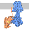
| Title | Cryo-EM structure of Gi coupled Sphingosine 1-phosphate receptor bound with SAR247799 | ||||||||||||||||||||||||
 Components Components |
| ||||||||||||||||||||||||
 Keywords Keywords | SIGNALING PROTEIN / GPCR / S1PR1 | ||||||||||||||||||||||||
| Function / homology |  Function and homology information Function and homology informationcardiac muscle tissue growth involved in heart morphogenesis / blood vessel maturation / sphingolipid binding / sphingosine-1-phosphate receptor activity / Lysosphingolipid and LPA receptors / endothelial cell differentiation / heart trabecula morphogenesis / regulation of bone mineralization / negative regulation of stress fiber assembly / sphingosine-1-phosphate receptor signaling pathway ...cardiac muscle tissue growth involved in heart morphogenesis / blood vessel maturation / sphingolipid binding / sphingosine-1-phosphate receptor activity / Lysosphingolipid and LPA receptors / endothelial cell differentiation / heart trabecula morphogenesis / regulation of bone mineralization / negative regulation of stress fiber assembly / sphingosine-1-phosphate receptor signaling pathway / positive regulation of positive chemotaxis / regulation of bone resorption / lamellipodium assembly / transmission of nerve impulse / regulation of eating behavior / adenylate cyclase inhibitor activity / T cell migration / positive regulation of protein localization to cell cortex / positive regulation of relaxation of smooth muscle / Adenylate cyclase inhibitory pathway / positive regulation of smooth muscle cell proliferation / D2 dopamine receptor binding / adenylate cyclase-inhibiting serotonin receptor signaling pathway / G protein-coupled serotonin receptor binding / cellular response to forskolin / regulation of mitotic spindle organization / mast cell degranulation / chemokine-mediated signaling pathway / electron transport chain / neuropeptide signaling pathway / Regulation of insulin secretion / response to prostaglandin E / positive regulation of cholesterol biosynthetic process / G protein-coupled receptor binding / response to peptide hormone / chemotaxis / G protein-coupled receptor activity / G-protein beta/gamma-subunit complex binding / adenylate cyclase-inhibiting G protein-coupled receptor signaling pathway / adenylate cyclase-modulating G protein-coupled receptor signaling pathway / phospholipase C-activating G protein-coupled receptor signaling pathway / Olfactory Signaling Pathway / Activation of the phototransduction cascade / G protein-coupled acetylcholine receptor signaling pathway / G beta:gamma signalling through PLC beta / Presynaptic function of Kainate receptors / Thromboxane signalling through TP receptor / actin cytoskeleton organization / Activation of G protein gated Potassium channels / Inhibition of voltage gated Ca2+ channels via Gbeta/gamma subunits / G-protein activation / Glucagon signaling in metabolic regulation / G beta:gamma signalling through CDC42 / Prostacyclin signalling through prostacyclin receptor / Synthesis, secretion, and inactivation of Glucagon-like Peptide-1 (GLP-1) / GDP binding / photoreceptor disc membrane / G beta:gamma signalling through BTK / ADP signalling through P2Y purinoceptor 12 / Glucagon-type ligand receptors / Sensory perception of sweet, bitter, and umami (glutamate) taste / Adrenaline,noradrenaline inhibits insulin secretion / neuron differentiation / Vasopressin regulates renal water homeostasis via Aquaporins / G alpha (z) signalling events / Glucagon-like Peptide-1 (GLP1) regulates insulin secretion / ADP signalling through P2Y purinoceptor 1 / cellular response to catecholamine stimulus / cell migration / G beta:gamma signalling through PI3Kgamma / ADORA2B mediated anti-inflammatory cytokines production / adenylate cyclase-activating dopamine receptor signaling pathway / cellular response to prostaglandin E stimulus / Cooperation of PDCL (PhLP1) and TRiC/CCT in G-protein beta folding / presynapse / GPER1 signaling / heterotrimeric G-protein complex / Inactivation, recovery and regulation of the phototransduction cascade / G alpha (12/13) signalling events / G-protein beta-subunit binding / angiogenesis / extracellular vesicle / Thrombin signalling through proteinase activated receptors (PARs) / adenylate cyclase-activating G protein-coupled receptor signaling pathway / signaling receptor complex adaptor activity / ciliary basal body / Interleukin-4 and Interleukin-13 signaling / GTPase binding / midbody / G protein activity / Ca2+ pathway / cell cortex / High laminar flow shear stress activates signaling by PIEZO1 and PECAM1:CDH5:KDR in endothelial cells / G alpha (i) signalling events / Potential therapeutics for SARS / G alpha (s) signalling events / G alpha (q) signalling events / Hydrolases; Acting on acid anhydrides; Acting on GTP to facilitate cellular and subcellular movement / Ras protein signal transduction / electron transfer activity Similarity search - Function | ||||||||||||||||||||||||
| Biological species |  Homo sapiens (human) Homo sapiens (human)  Mus musculus (house mouse) Mus musculus (house mouse)  Escherichia coli (E. coli) Escherichia coli (E. coli) | ||||||||||||||||||||||||
| Method | ELECTRON MICROSCOPY / single particle reconstruction / cryo EM / Resolution: 2.97 Å | ||||||||||||||||||||||||
 Authors Authors | Yu, L.Y. / Jiao, H.Z. / Ti, R.J. / Pang, B. / Gan, B. / Ren, R.B. | ||||||||||||||||||||||||
| Funding support |  China, 1items China, 1items
| ||||||||||||||||||||||||
 Citation Citation | Journal: PLoS Biol / Year: 2026 Title: Structural insights into subtype-specific agonist recognition by sphingosine-1-phosphate receptors. Authors: Leiye Yu / Haizhan Jiao / Bin Pang / Rujuan Ti / Bing Gan / Zhaoyang Qin / Jinxin Wang / Lizhe Zhu / Hongli Hu / Ruobing Ren / Abstract: Sphingosine-1-phosphate (S1P), a key metabolite of sphingolipids, plays crucial roles in a wide range of physiological and pathological processes. S1P primarily exerts its functions by binding to G ...Sphingosine-1-phosphate (S1P), a key metabolite of sphingolipids, plays crucial roles in a wide range of physiological and pathological processes. S1P primarily exerts its functions by binding to G protein-coupled sphingosine-1-phosphate receptors (S1PRs), which comprise five subtypes (S1PR1-5) in humans, thereby activating these receptors and their downstream signaling pathways. Understanding the molecular determinants that govern agonist selectivity among different S1PR subtypes is vital for the rational and precise development of targeted therapeutic agents. Here, four cryo-electron microscopy structures of agonist-bound S1PR1-Gi1 complexes are reported. Through an integrated approach combining structural analysis, molecular dynamics simulations, and pharmacological assays, the molecular basis for the selectivity of CYM5442, HY-X-1011, Ponesimod, and SAR247799 toward S1PR1 over S1PR2-S1PR5 is uncovered. Nonconserved residues within the ligand-binding pocket and at the Gi1-protein interface contribute to S1PR1 selectivity by these agonists. A distinct agonist binding orientation toward transmembrane helices 5-7, combined with branched substituents that increase the agonist's molecular width, results in steric clashes with residues in S1PR3. Additionally, branched moieties located at the tail portions of the agonist restrict its deep insertion into the binding pocket of both S1PR3 and S1PR5. These structural features collectively enhance its selectivity for S1PR1 over S1PR3 and S1PR5. Furthermore, polar interactions with conserved polar residues in the top region of the binding pocket also influence agonist selectivity. Besides, the relatively broad molecular width of the agonist sterically hinders its binding into S1PR2 and S1PR4 pocket by nonconserved residue pairs bearing bulky side chains. These findings establish a structural framework for the rational design of next-generation S1PR1 highly selective agonists with improved therapeutic potential. | ||||||||||||||||||||||||
| History |
|
- Structure visualization
| Structure viewer | Molecule: MolmilJmol/JSmol |
|---|
- Downloads & links
-Download
| PDBx/mmCIF format | 9vo1.cif.gz | 246.6 KB | Display | PDBx/mmCIF format |
|---|---|---|---|---|
| PDB format | pdb9vo1.ent.gz | Display | PDB format | |
| PDBx/mmJSON format | 9vo1.json.gz | Tree view | PDBx/mmJSON format | |
| Others |  Other downloads Other downloads |
-Validation report
| Arichive directory | https://data.pdbj.org/pub/pdb/validation_reports/vo/9vo1ftp://data.pdbj.org/pub/pdb/validation_reports/vo/9vo1 | HTTPS FTP |
|---|
-Related structure data
| Related structure data |  65221MC  9vnyC  9vnzC  9vo0C M: map data used to model this data C: citing same article ( |
|---|---|
| Similar structure data |


-Links
 PDBj
PDBj


















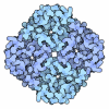





- Assembly
| Deposited unit | 
|
|---|---|
| 1 |
|
-Components
-Guanine nucleotide-binding protein ... , 3 types, 3 molecules ACD
| #1: Protein | Mass: 39286.891 Da / Num. of mol.: 1 Source method: isolated from a genetically manipulated source Source: (gene. exp.) Homo sapiens (human) / Gene: GNB1 / Cell line (production host): Sf9 / Production host:   Spodoptera frugiperda (fall armyworm) / References: UniProt: P62873 Spodoptera frugiperda (fall armyworm) / References: UniProt: P62873 |
|---|---|
| #2: Protein | Mass: 7861.143 Da / Num. of mol.: 1 Source method: isolated from a genetically manipulated source Source: (gene. exp.) Homo sapiens (human) / Gene: GNG2 / Cell line (production host): Sf9 / Production host: Spodoptera frugiperda (fall armyworm) / References: UniProt: P59768 |
| #5: Protein | Mass: 40415.031 Da / Num. of mol.: 1 Source method: isolated from a genetically manipulated source Source: (gene. exp.) Homo sapiens (human) / Gene: GNAI1 / Cell line (production host): Sf9 / Production host: Spodoptera frugiperda (fall armyworm)References: UniProt: P63096, Hydrolases; Acting on acid anhydrides; Acting on GTP to facilitate cellular and subcellular movement |
-Antibody / Protein / Non-polymers , 3 types, 3 molecules EF
| #3: Antibody | Mass: 26679.721 Da / Num. of mol.: 1 Source method: isolated from a genetically manipulated source Source: (gene. exp.) Mus musculus (house mouse) / Cell line (production host): Hi5 / Production host: Trichoplusia ni (cabbage looper) |
|---|---|
| #4: Protein | Mass: 60092.078 Da / Num. of mol.: 1 Source method: isolated from a genetically manipulated source Details: Author remarks: Molecule 5 (chain F) is a fusion protein. 1-16 is the HA signal sequence, 17-24 is flag tag sequence,25-32 is the sequence of HRV 3C enzyme cutting site, 33-34 is two linker ...Details: Author remarks: Molecule 5 (chain F) is a fusion protein. 1-16 is the HA signal sequence, 17-24 is flag tag sequence,25-32 is the sequence of HRV 3C enzyme cutting site, 33-34 is two linker amino acids and 35-140, is the sequence of Soluble cytochrome b562, UniProt ID P0ABE7. 523-524 is two linker amino acids and 525-534 is the sequence of His tag. Source: (gene. exp.) Escherichia coli (E. coli), (gene. exp.) Homo sapiens (human)Gene: cybC, S1PR1, CHEDG1, EDG1 / Cell line (production host): Sf9 / Production host: Spodoptera frugiperda (fall armyworm) / References: UniProt: P0ABE7, UniProt: P21453 |
| #6: Chemical | ChemComp-A1LYQ / Mass: 425.822 Da / Num. of mol.: 1 / Source method: obtained synthetically / Formula: C21H16ClN3O5 / Feature type: SUBJECT OF INVESTIGATION |
-Details
| Has ligand of interest | Y |
|---|---|
| Has protein modification | Y |
-Experimental details
-Experiment
| Experiment | Method: ELECTRON MICROSCOPY |
|---|---|
| EM experiment | Aggregation state: PARTICLE / 3D reconstruction method: single particle reconstruction |
- Sample preparation
| Component |
| ||||||||||||||||||||||||
|---|---|---|---|---|---|---|---|---|---|---|---|---|---|---|---|---|---|---|---|---|---|---|---|---|---|
| Source (natural) |
| ||||||||||||||||||||||||
| Source (recombinant) |
| ||||||||||||||||||||||||
| Buffer solution | pH: 7.5 | ||||||||||||||||||||||||
| Specimen | Embedding applied: NO / Shadowing applied: NO / Staining applied: NO / Vitrification applied: YES | ||||||||||||||||||||||||
| Vitrification | Cryogen name: ETHANE |
- Electron microscopy imaging
| Experimental equipment |  Model: Titan Krios / Image courtesy: FEI Company |
|---|---|
| Microscopy | Model: TFS KRIOS |
| Electron gun | Electron source:  FIELD EMISSION GUN / Accelerating voltage: 300 kV / Illumination mode: FLOOD BEAM FIELD EMISSION GUN / Accelerating voltage: 300 kV / Illumination mode: FLOOD BEAM |
| Electron lens | Mode: BRIGHT FIELD / Nominal defocus max: 1900 nm / Nominal defocus min: 1100 nm |
| Image recording | Electron dose: 56.14 e/Å2 / Film or detector model: GATAN K3 (6k x 4k) |
- Processing
| EM software |
| ||||||||||||||||||||||||
|---|---|---|---|---|---|---|---|---|---|---|---|---|---|---|---|---|---|---|---|---|---|---|---|---|---|
| CTF correction | Type: NONE | ||||||||||||||||||||||||
| 3D reconstruction | Resolution: 2.97 Å / Resolution method: FSC 0.143 CUT-OFF / Num. of particles: 94216 / Symmetry type: POINT | ||||||||||||||||||||||||
| Refinement | Highest resolution: 2.97 Å Stereochemistry target values: REAL-SPACE (WEIGHTED MAP SUM AT ATOM CENTERS) | ||||||||||||||||||||||||
| Refine LS restraints |
|
