Journal: Nat Commun / Year: 2026 Title: Two types of sheathed archaellum structures from Haloarcula marismortui differ in their outer layer architectures. Authors: Vladimir A Meshcheryakov / Jaekyung Hyun / Satoshi Shibata / Alexey S Syutkin / Mikhail G Pyatibratov / Matthias Wolf / Abstract: Many archaea swim by means of rotating helical filaments called archaella. Most archaella are about 10 nm in diameter and comprise multiple copies of the protein archaellin. Here, we describe two ...Many archaea swim by means of rotating helical filaments called archaella. Most archaella are about 10 nm in diameter and comprise multiple copies of the protein archaellin. Here, we describe two archaellum structures formed by the ArlA2 or ArlB archaellins from the haloarchaeon Haloarcula marismortui. We found that both filaments have an additional proteinaceous outer sheath surrounding their inner core, a feature not observed previously in archaea. The outer sheath structures of the two filaments differ fundamentally. The outer domain of ArlB archaellin rotates by 180, forming stable dimers that likely increase the filament rigidity. In contrast, neither rotation nor dimerization of the outer domain was observed in ArlA2 filaments. 3D variability analysis demonstrated that the motions of ArlA2 and ArlB filaments are significantly distinct. Additionally, the ArlB filament displays a larger negatively charged surface area than ArlA2, which may help adaptation to higher salt concentrations. Our structural findings provide insight into how the two filaments can adapt to their different environments. Larger archaellins with additional outer domains can be found in various groups of archaea. We propose that such proteins may allow hosts to modify the properties of their archaella to enhance environmental adaptation.
History
Deposition
May 30, 2025
Deposition site: PDBJ / Processing site: PDBJ
Revision 1.0
May 27, 2026
Provider: repository / Type: Initial release
Revision 1.0
May 27, 2026
Data content type: EM metadata / Data content type: EM metadata / Provider: repository / Type: Initial release
Revision 1.0
May 27, 2026
Data content type: Additional map / Part number: 1 / Data content type: Additional map / Provider: repository / Type: Initial release
Revision 1.0
May 27, 2026
Data content type: FSC / Data content type: FSC / Provider: repository / Type: Initial release
Revision 1.0
May 27, 2026
Data content type: Half map / Part number: 1 / Data content type: Half map / Provider: repository / Type: Initial release
Revision 1.0
May 27, 2026
Data content type: Half map / Part number: 2 / Data content type: Half map / Provider: repository / Type: Initial release
Revision 1.0
May 27, 2026
Data content type: Image / Data content type: Image / Provider: repository / Type: Initial release
Revision 1.0
May 27, 2026
Data content type: Primary map / Data content type: Primary map / Provider: repository / Type: Initial release
Data content type: EM metadata / Data content type: EM metadata / EM metadata / Group: Database references / Experimental summary / Data content type: EM metadata / EM metadata / Category: citation / em_admin / Data content type: EM metadata / EM metadata / Item: _citation.journal_volume / _em_admin.last_update
In the structure databanks used in Yorodumi, some data are registered as the other names, "COVID-19 virus" and "2019-nCoV". Here are the details of the virus and the list of structure data.
Jan 31, 2019. EMDB accession codes are about to change! (news from PDBe EMDB page)
EMDB accession codes are about to change! (news from PDBe EMDB page)
The allocation of 4 digits for EMDB accession codes will soon come to an end. Whilst these codes will remain in use, new EMDB accession codes will include an additional digit and will expand incrementally as the available range of codes is exhausted. The current 4-digit format prefixed with “EMD-” (i.e. EMD-XXXX) will advance to a 5-digit format (i.e. EMD-XXXXX), and so on. It is currently estimated that the 4-digit codes will be depleted around Spring 2019, at which point the 5-digit format will come into force.
The EM Navigator/Yorodumi systems omit the EMD- prefix.
Related info.:Q: What is EMD? / ID/Accession-code notation in Yorodumi/EM Navigator
Yorodumi is a browser for structure data from EMDB, PDB, SASBDB, etc.
This page is also the successor to EM Navigator detail page, and also detail information page/front-end page for Omokage search.
The word "yorodu" (or yorozu) is an old Japanese word meaning "ten thousand". "mi" (miru) is to see.
Related info.:EMDB / PDB / SASBDB / Comparison of 3 databanks / Yorodumi Search / Aug 31, 2016. New EM Navigator & Yorodumi / Yorodumi Papers / Jmol/JSmol / Function and homology information / Changes in new EM Navigator and Yorodumi
 Movie
Movie Controller


 Open data
 Basic information
 Components
 Keywords
 Function and homology information
 Haloarcula marismortui (Halophile)
 Authors
 Japan, 1items
  Citation

 Structure visualization
 Downloads & links
 Other downloads


 PDBj
PDBj Assembly

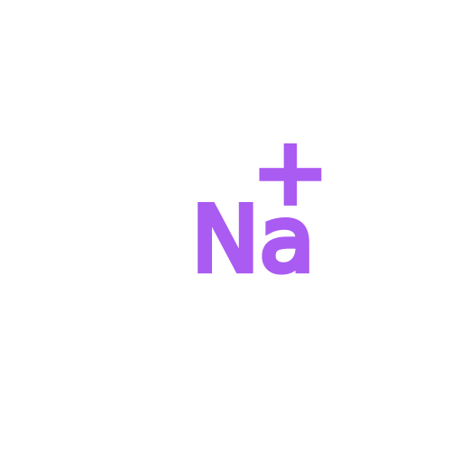 Mass: 22.990 Da / Num. of mol.: 33 / Source method: obtained synthetically / Formula: Na
 Sample preparation
 Electron microscopy imaging
 FIELD EMISSION GUN / Accelerating voltage: 300 kV / Illumination mode: FLOOD BEAM
 Processing