 Movie
Movie Controller


+ Open data

- Basic information
| Entry | Database: PDB / ID: 9sfw | |||||||||
|---|---|---|---|---|---|---|---|---|---|---|
| Title | Structure of Pex8 in complex with peroxisomal receptor Pex5 | |||||||||
 Components Components |
| |||||||||
 Keywords Keywords | PROTEIN TRANSPORT / Peroxisomal translocation receptor / HEAT Repeat | |||||||||
| Function / homology |  Function and homology information Function and homology informationperoxisome matrix targeting signal-1 binding / protein import into peroxisome matrix, docking / peroxisomal membrane / peroxisomal matrix / cytosol Similarity search - Function | |||||||||
| Biological species |  Komagataella pastoris (fungus) Komagataella pastoris (fungus) | |||||||||
| Method | ELECTRON MICROSCOPY / single particle reconstruction / cryo EM / Resolution: 4.5 Å | |||||||||
 Authors Authors | Ekal, L. / Mullapudi, E. / Chojnowski, G. / Wilmanns, M. | |||||||||
| Funding support |  Germany, 1items Germany, 1items
| |||||||||
 Citation Citation | Journal: To Be Published Title: Cryo-EM structure of Pichia pastoris Pex8-Pex5 receptor complex Authors: Ekal, L. / Mullapudi, E. / Chojnowski, G. / WIlmanns, M. | |||||||||
| History |
|
- Structure visualization
| Structure viewer | Molecule: MolmilJmol/JSmol |
|---|
- Downloads & links
-Download
| PDBx/mmCIF format | 9sfw.cif.gz | 215.3 KB | Display | PDBx/mmCIF format |
|---|---|---|---|---|
| PDB format | pdb9sfw.ent.gz | 164.3 KB | Display | PDB format |
| PDBx/mmJSON format | 9sfw.json.gz | Tree view | PDBx/mmJSON format | |
| Others |  Other downloads Other downloads |
-Validation report
| Arichive directory | https://data.pdbj.org/pub/pdb/validation_reports/sf/9sfwftp://data.pdbj.org/pub/pdb/validation_reports/sf/9sfw | HTTPS FTP |
|---|
-Related structure data
| Related structure data |  54867MC M: map data used to model this data C: citing same article ( |
|---|---|
| Similar structure data |
-Links
 PDBj
PDBj
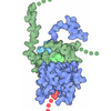

- Assembly
| Deposited unit | 
|
|---|---|
| 1 |
|
-Components
| #1: Protein | Mass: 81081.195 Da / Num. of mol.: 1 Source method: isolated from a genetically manipulated source Details: Pex8 construct missing 1-33aa was used for affinity purification and subsequent experimental analysis. Source: (gene. exp.) Komagataella pastoris (fungus) / Gene: PEX8, PER3 / Production host:   Escherichia coli BL21(DE3) (bacteria) / Strain (production host): Lobstr / References: UniProt: Q01962 Escherichia coli BL21(DE3) (bacteria) / Strain (production host): Lobstr / References: UniProt: Q01962 |
|---|---|
| #2: Protein | Mass: 65143.129 Da / Num. of mol.: 1 Source method: isolated from a genetically manipulated source Details: The mismatch highlighted in the alignment is due to a small peptide in the structure, spanning residues 54-66, which is aligning incorrectly at the region starting with residue 243 in the sample sequence. Source: (gene. exp.) Komagataella pastoris (fungus) / Gene: PEX5, PAS8 / Production host: Escherichia coli BL21(DE3) (bacteria) / Strain (production host): BL21-CodonPlus-RIL / References: UniProt: P33292 |
| Has protein modification | N |
-Experimental details
-Experiment
| Experiment | Method: ELECTRON MICROSCOPY |
|---|---|
| EM experiment | Aggregation state: PARTICLE / 3D reconstruction method: single particle reconstruction |
- Sample preparation
| Component | Name: Pex8-Pex5 Complex / Type: COMPLEX / Entity ID: all / Source: RECOMBINANT | ||||||||||||||||||||
|---|---|---|---|---|---|---|---|---|---|---|---|---|---|---|---|---|---|---|---|---|---|
| Molecular weight | Value: 0.143 MDa / Experimental value: NO | ||||||||||||||||||||
| Source (natural) | Organism: Komagataella pastoris (fungus) | ||||||||||||||||||||
| Source (recombinant) | Organism: Escherichia coli BL21(DE3) (bacteria) | ||||||||||||||||||||
| Buffer solution | pH: 7.5 | ||||||||||||||||||||
| Buffer component |
| ||||||||||||||||||||
| Specimen | Conc.: 1 mg/ml / Embedding applied: NO / Shadowing applied: NO / Staining applied: NO / Vitrification applied: YES | ||||||||||||||||||||
| Specimen support | Grid material: COPPER / Grid mesh size: 300 divisions/in. / Grid type: Quantifoil R2/1 | ||||||||||||||||||||
| Vitrification | Instrument: FEI VITROBOT MARK IV / Cryogen name: ETHANE-PROPANE / Humidity: 100 % / Chamber temperature: 279 K |
- Electron microscopy imaging
| Experimental equipment |  Model: Titan Krios / Image courtesy: FEI Company |
|---|---|
| Microscopy | Model: TFS KRIOS |
| Electron gun | Electron source:  FIELD EMISSION GUN / Accelerating voltage: 300 kV / Illumination mode: FLOOD BEAM FIELD EMISSION GUN / Accelerating voltage: 300 kV / Illumination mode: FLOOD BEAM |
| Electron lens | Mode: BRIGHT FIELD / Nominal magnification: 130000 X / Nominal defocus max: 2500 nm / Nominal defocus min: 750 nm / Alignment procedure: COMA FREE |
| Specimen holder | Cryogen: NITROGEN / Specimen holder model: FEI TITAN KRIOS AUTOGRID HOLDER |
| Image recording | Electron dose: 64.5 e/Å2 / Film or detector model: GATAN K3 (6k x 4k) |
- Processing
| EM software |
| |||||||||||||||||||||||||||||||||||||||||||||||||||||||||||||||||||||||||||||||||||||||||||||||||||||||||||
|---|---|---|---|---|---|---|---|---|---|---|---|---|---|---|---|---|---|---|---|---|---|---|---|---|---|---|---|---|---|---|---|---|---|---|---|---|---|---|---|---|---|---|---|---|---|---|---|---|---|---|---|---|---|---|---|---|---|---|---|---|---|---|---|---|---|---|---|---|---|---|---|---|---|---|---|---|---|---|---|---|---|---|---|---|---|---|---|---|---|---|---|---|---|---|---|---|---|---|---|---|---|---|---|---|---|---|---|---|
| CTF correction | Type: NONE | |||||||||||||||||||||||||||||||||||||||||||||||||||||||||||||||||||||||||||||||||||||||||||||||||||||||||||
| Symmetry | Point symmetry: C1 (asymmetric) | |||||||||||||||||||||||||||||||||||||||||||||||||||||||||||||||||||||||||||||||||||||||||||||||||||||||||||
| 3D reconstruction | Resolution: 4.5 Å / Resolution method: FSC 0.143 CUT-OFF / Num. of particles: 85869 / Algorithm: FOURIER SPACE / Symmetry type: POINT | |||||||||||||||||||||||||||||||||||||||||||||||||||||||||||||||||||||||||||||||||||||||||||||||||||||||||||
| Atomic model building | Protocol: RIGID BODY FIT | |||||||||||||||||||||||||||||||||||||||||||||||||||||||||||||||||||||||||||||||||||||||||||||||||||||||||||
| Atomic model building | Details: version 3 / Source name: AlphaFold / Type: in silico model | |||||||||||||||||||||||||||||||||||||||||||||||||||||||||||||||||||||||||||||||||||||||||||||||||||||||||||
| Refinement | Resolution: 4.5→4.5 Å / Cor.coef. Fo:Fc: 0.926 / WRfactor Rwork: 0.389 / SU B: 34.783 / SU ML: 0.349 / Average fsc free: 0 / Average fsc overall: 0.9156 / Average fsc work: 0.9156 Details: Hydrogens have been added in their riding positions
| |||||||||||||||||||||||||||||||||||||||||||||||||||||||||||||||||||||||||||||||||||||||||||||||||||||||||||
| Solvent computation | Solvent model: NONE | |||||||||||||||||||||||||||||||||||||||||||||||||||||||||||||||||||||||||||||||||||||||||||||||||||||||||||
| Displacement parameters | Biso mean: 459.989 Å2 | |||||||||||||||||||||||||||||||||||||||||||||||||||||||||||||||||||||||||||||||||||||||||||||||||||||||||||
| Refine LS restraints |
|