 Movie
Movie Controller


+ Open data

- Basic information
| Entry | Database: PDB / ID: 9pvm | |||||||||
|---|---|---|---|---|---|---|---|---|---|---|
| Title | cryoEM structure of drug bound human SLC33A1 transporter | |||||||||
 Components Components | Acetyl-coenzyme A transporter 1 | |||||||||
 Keywords Keywords | MEMBRANE PROTEIN / solute carrier / ER membrane transporter / cryoEM / drug binding | |||||||||
| Function / homology |  Function and homology information Function and homology informationacetyl-CoA transmembrane transporter activity / acetyl-CoA transmembrane transport / Defective SLC33A1 causes spastic paraplegia 42 (SPG42) / Transport of vitamins, nucleosides, and related molecules / transmembrane transport / Golgi membrane / endoplasmic reticulum membrane / protein homodimerization activity / membrane / plasma membrane Similarity search - Function | |||||||||
| Biological species |  Homo sapiens (human) Homo sapiens (human) | |||||||||
| Method | ELECTRON MICROSCOPY / single particle reconstruction / cryo EM / Resolution: 3.27 Å | |||||||||
 Authors Authors | Rafiq, M. / Lander, G.C. | |||||||||
| Funding support |  United States, 1items United States, 1items
| |||||||||
 Citation Citation | Journal: bioRxiv / Year: 2026 Title: Pharmacological Inhibition of SLC33A1 Promotes Endoplasmic Reticulum Hyperoxidation and Induces Adaptive IRE1/XBP1s Signaling. Authors: Sergei Kutseikin / Maria Rafiq / Prerona Bora / Shanshan Liu / Rick A Homan / Jeffrey T Mindrebo / Matthew Holcomb / H Michael Petrassi / Huang Qiu / Anastasiya Redkina / Justyna Sosna / ...Authors: Sergei Kutseikin / Maria Rafiq / Prerona Bora / Shanshan Liu / Rick A Homan / Jeffrey T Mindrebo / Matthew Holcomb / H Michael Petrassi / Huang Qiu / Anastasiya Redkina / Justyna Sosna / Thanh-Trang Lee / Xiao Hu / Stefano Forli / Christopher G Parker / Gabriel C Lander / Kivanc Birsoy / Enrique Saez / R Luke Wiseman / Abstract: The endoplasmic reticulum (ER) transporter solute carrier family 33 member 1 (SLC33A1) has emerged as an attractive therapeutic target in etiologically diverse diseases, ranging from lung cancer to ...The endoplasmic reticulum (ER) transporter solute carrier family 33 member 1 (SLC33A1) has emerged as an attractive therapeutic target in etiologically diverse diseases, ranging from lung cancer to neurodegenerative disorders. Yet, no pharmacologic SLC33A1 modulators have been described. Here, we show that the small molecule IXA4, a highly selective activator of the adaptive IRE1/XBP1s signaling arm of the unfolded protein response (UPR), binds to SLC33A1 and inhibits its activity. Genetic depletion of phenocopies the selective induction of IRE1/XBP1s signaling brought about by IXA4 treatment. Chemoproteomic analyses and cryo-electron microscopy show that IXA4 binds SLC33A1 within the central channel to inhibit transport of its substrate metabolite(s). Binding of IXA4 to SLC33A1 leads to the accumulation of oxidized glutathione within the ER, hyperoxidizing the ER lumen and inducing activation of adaptive IRE1/XBP1s signaling. Consistent with this function, we find that pharmacologic inhibition of SLC33A1 with IXA4 selectively reduces viability of KEAP1-deficient lung adenocarcinoma cells that have elevated levels of glutathione, mimicking the sensitivity of these cells to genetic deletion of SLC33A1. Our work demonstrates a new physiologic role of SLC33A1 in regulation of ER redox homeostasis and designates IXA4 as a pharmacologic inhibitor of SLC33A1 that can be used to evaluate the biological impact and therapeutic utility of SLC33A1 inhibition in homeostasis and in disease. | |||||||||
| History |
|
- Structure visualization
| Structure viewer | Molecule: MolmilJmol/JSmol |
|---|
- Downloads & links
-Download
| PDBx/mmCIF format | 9pvm.cif.gz | 189.2 KB | Display | PDBx/mmCIF format |
|---|---|---|---|---|
| PDB format | pdb9pvm.ent.gz | Display | PDB format | |
| PDBx/mmJSON format | 9pvm.json.gz | Tree view | PDBx/mmJSON format | |
| Others |  Other downloads Other downloads |
-Validation report
| Arichive directory | https://data.pdbj.org/pub/pdb/validation_reports/pv/9pvmftp://data.pdbj.org/pub/pdb/validation_reports/pv/9pvm | HTTPS FTP |
|---|
-Related structure data
| Related structure data |  71893MC  9z5iC M: map data used to model this data C: citing same article ( |
|---|---|
| Similar structure data |

-Links
 PDBj
PDBj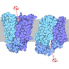

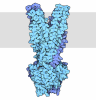
- Assembly
| Deposited unit | 
|
|---|---|
| 1 |
|
-Components
| #1: Protein | Mass: 64376.145 Da / Num. of mol.: 1 / Mutation: no Source method: isolated from a genetically manipulated source Details: Wild type, full length SLC33A11 / Source: (gene. exp.) Homo sapiens (human) / Cell line: Expi293 GnTI- / Gene: SLC33A1, ACATN, AT1 / Plasmid: pCDNA3.1Details (production host): Full-length human SLC33A1 cloned into pCDNA3.1 for expression in Expi293 GnTI- cells Cell (production host): Expi293 / Cell line (production host): Expi293 / Production host: Homo sapiens (human) / Tissue (production host): embryonic kidney / Variant (production host): GnTI- / References: UniProt: O00400 | ||||
|---|---|---|---|---|---|
| #2: Chemical | ChemComp-A1CTY / Mass: 436.504 Da / Num. of mol.: 1 / Source method: obtained synthetically / Formula: C24H28N4O4 / Feature type: SUBJECT OF INVESTIGATION | ||||
| #3: Chemical | 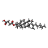 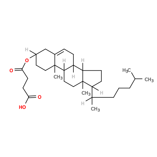 Mass: 486.726 Da / Num. of mol.: 2 / Source method: obtained synthetically / Formula: C31H50O4 Mass: 486.726 Da / Num. of mol.: 2 / Source method: obtained synthetically / Formula: C31H50O4Has ligand of interest | Y | Has protein modification | N | |
-Experimental details
-Experiment
| Experiment | Method: ELECTRON MICROSCOPY |
|---|---|
| EM experiment | Aggregation state: PARTICLE / 3D reconstruction method: single particle reconstruction |
- Sample preparation
| Component | Name: Full length SLC33A1 with ligand bound / Type: COMPLEX Details: Reconstituted into MSP1D1 and (POPC:DOPE:PS)lipid used Entity ID: #1 / Source: RECOMBINANT | |||||||||||||||
|---|---|---|---|---|---|---|---|---|---|---|---|---|---|---|---|---|
| Molecular weight | Value: 64.3 kDa/nm / Experimental value: NO | |||||||||||||||
| Source (natural) | Organism: Homo sapiens (human) / Cellular location: Endoplasmic reticulum | |||||||||||||||
| Source (recombinant) | Organism: Homo sapiens (human) / Cell: Expi293 GnTI- / Plasmid: pcDNA3.1 | |||||||||||||||
| Buffer solution | pH: 8 / Details: 150mM NaCl, 20mM Tris pH 8.0 | |||||||||||||||
| Buffer component |
| |||||||||||||||
| Specimen | Conc.: 5 mg/ml / Embedding applied: NO / Shadowing applied: NO / Staining applied: NO / Vitrification applied: YES Details: Protin was solubilized in DDM-CHS, buffer exchanged to LMNG-CHS and reconstituted into nanodisc using MSP1D1 and lipids POPC DOPE PS | |||||||||||||||
| Specimen support | Grid material: GOLD / Grid mesh size: 300 divisions/in. / Grid type: UltrAuFoil R0./1 | |||||||||||||||
| Vitrification | Instrument: HOMEMADE PLUNGER / Cryogen name: ETHANE / Humidity: 95 % / Chamber temperature: 277.15 K |
- Electron microscopy imaging
| Experimental equipment | 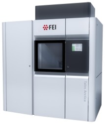 Model: Talos Arctica / Image courtesy: FEI Company |
|---|---|
| Microscopy | Model: FEI TALOS ARCTICA |
| Electron gun | Electron source:  FIELD EMISSION GUN / Accelerating voltage: 200 kV / Illumination mode: FLOOD BEAM FIELD EMISSION GUN / Accelerating voltage: 200 kV / Illumination mode: FLOOD BEAM |
| Electron lens | Mode: BRIGHT FIELD / Nominal magnification: 190000 X / Calibrated magnification: 189189 X / Nominal defocus max: 2000 nm / Nominal defocus min: 800 nm / Cs: 2.7 mm / C2 aperture diameter: 30 µm / Alignment procedure: COMA FREE |
| Specimen holder | Cryogen: NITROGEN / Specimen holder model: FEI TITAN KRIOS AUTOGRID HOLDER / Temperature (max): 77 K / Temperature (min): 70 K |
| Image recording | Average exposure time: 3.29 sec. / Electron dose: 40 e/Å2 / Film or detector model: TFS FALCON 4i (4k x 4k) / Num. of grids imaged: 4 / Num. of real images: 6468 |
| Image scans | Sampling size: 14 µm / Width: 4096 / Height: 4096 |
- Processing
| EM software |
| ||||||||||||||||||||||||||||||||||||
|---|---|---|---|---|---|---|---|---|---|---|---|---|---|---|---|---|---|---|---|---|---|---|---|---|---|---|---|---|---|---|---|---|---|---|---|---|---|
| CTF correction | Type: PHASE FLIPPING ONLY | ||||||||||||||||||||||||||||||||||||
| Particle selection | Num. of particles selected: 3119666 | ||||||||||||||||||||||||||||||||||||
| Symmetry | Point symmetry: C1 (asymmetric) | ||||||||||||||||||||||||||||||||||||
| 3D reconstruction | Resolution: 3.27 Å / Resolution method: FSC 0.143 CUT-OFF / Num. of particles: 457520 / Algorithm: FOURIER SPACE / Num. of class averages: 1 / Symmetry type: POINT | ||||||||||||||||||||||||||||||||||||
| Atomic model building | B value: 180 / Protocol: RIGID BODY FIT | ||||||||||||||||||||||||||||||||||||
| Atomic model building | Source name: AlphaFold / Type: in silico model | ||||||||||||||||||||||||||||||||||||
| Refinement | Highest resolution: 3.27 Å Stereochemistry target values: REAL-SPACE (WEIGHTED MAP SUM AT ATOM CENTERS) | ||||||||||||||||||||||||||||||||||||
| Refine LS restraints |
|
