 Movie
Movie Controller


+ Open data

- Basic information
| Entry | Database: PDB / ID: 9nqy | ||||||||||||
|---|---|---|---|---|---|---|---|---|---|---|---|---|---|
| Title | Nipah Virus Matrix Lattice in Complex with Human Cell Membrane | ||||||||||||
 Components Components | Matrix protein | ||||||||||||
 Keywords Keywords | VIRUS LIKE PARTICLE / Assembly Lattice | ||||||||||||
| Function / homology |  Function and homology information Function and homology informationvirion assembly / virion component / structural constituent of virion / host cell cytoplasm / host cell nucleus / host cell plasma membrane Similarity search - Function | ||||||||||||
| Biological species |  Henipavirus nipahense Henipavirus nipahense | ||||||||||||
| Method | ELECTRON MICROSCOPY / subtomogram averaging / cryo EM / Resolution: 7 Å | ||||||||||||
 Authors Authors | Upadhye, V.V. / Dick, R.A. | ||||||||||||
| Funding support |  United States, 3items United States, 3items
| ||||||||||||
 Citation Citation | Journal: bioRxiv / Year: 2026 Title: Cryo-electron tomography of Nipah virus structural protein complexes in virus-like particles. Authors: Viraj V Upadhye / Jean F Lee / Nihan Ercanli / Clifton Ricana / Amy C Hinsley / Martin Obr / Ludovic Autin / Florian K M Schur / Hector C Aguilar / Robert A Dick /   Abstract: Nipah virus (NiV) is a BSL-4 zoonotic paramyxovirus with ~75% human mortality. The matrix protein (M) of NiV and other paramyxoviruses binds the inner leaflet of the cellular plasma membrane, ...Nipah virus (NiV) is a BSL-4 zoonotic paramyxovirus with ~75% human mortality. The matrix protein (M) of NiV and other paramyxoviruses binds the inner leaflet of the cellular plasma membrane, orchestrating virion assembly by bringing together transmembrane glycoproteins (F/G) and ribonucleoprotein complexes (N). However, the interactions of these full-length proteins within membrane complexes remain elusive. Using cryo-electron tomography and subtomogram averaging of virus like particles (VLPs), we interrogated the protein:protein interactions of the main NiV structural proteins M/N/F/G. The M lattice structure determined to 7Å revealed a novel M-dimer arrangement that yielded two distinct repeating holes. Notably, F-trimers were arranged above only one of the two holes, dependent on F's cytoplasmic tail. G was enriched in regions of higher M-VLP curvature, while N dramatically increased M-VLP pleomorphism. This work provides novel insights into paramyxoviral protein complexes, structures, and morphology. | ||||||||||||
| History |
|
- Structure visualization
| Structure viewer | Molecule: MolmilJmol/JSmol |
|---|
- Downloads & links
-Download
| PDBx/mmCIF format | 9nqy.cif.gz | 824 KB | Display | PDBx/mmCIF format |
|---|---|---|---|---|
| PDB format | pdb9nqy.ent.gz | 584.4 KB | Display | PDB format |
| PDBx/mmJSON format | 9nqy.json.gz | Tree view | PDBx/mmJSON format | |
| Others |  Other downloads Other downloads |
-Validation report
| Arichive directory | https://data.pdbj.org/pub/pdb/validation_reports/nq/9nqyftp://data.pdbj.org/pub/pdb/validation_reports/nq/9nqy | HTTPS FTP |
|---|
-Related structure data
| Related structure data |  49696MC M: map data used to model this data C: citing same article ( |
|---|---|
| Similar structure data |
-Links
 PDBj
PDBj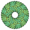
- Assembly
| Deposited unit | 
|
|---|---|
| 1 |
|
-Components
| #1: Protein | Mass: 44558.980 Da / Num. of mol.: 18 Source method: isolated from a genetically manipulated source Source: (gene. exp.) Henipavirus nipahense / Production host:  Homo sapiens (human) / References: UniProt: Q9IK90 Homo sapiens (human) / References: UniProt: Q9IK90Has protein modification | N | |
|---|
-Experimental details
-Experiment
| Experiment | Method: ELECTRON MICROSCOPY |
|---|---|
| EM experiment | Aggregation state: 3D ARRAY / 3D reconstruction method: subtomogram averaging |
- Sample preparation
| Component | Name: Henipavirus nipahense / Type: VIRUS / Entity ID: all / Source: RECOMBINANT | ||||||||||||||||
|---|---|---|---|---|---|---|---|---|---|---|---|---|---|---|---|---|---|
| Source (natural) | Organism: Henipavirus nipahense / Strain: Malaysia | ||||||||||||||||
| Source (recombinant) | Organism: Homo sapiens (human) | ||||||||||||||||
| Details of virus | Empty: YES / Enveloped: YES / Isolate: SPECIES / Type: VIRUS-LIKE PARTICLE | ||||||||||||||||
| Buffer solution | pH: 7.5 | ||||||||||||||||
| Buffer component |
| ||||||||||||||||
| Specimen | Embedding applied: NO / Shadowing applied: NO / Staining applied: NO / Vitrification applied: YES | ||||||||||||||||
| Specimen support | Grid material: GOLD / Grid mesh size: 300 divisions/in. / Grid type: Quantifoil R2/2 | ||||||||||||||||
| Vitrification | Instrument: LEICA EM GP / Cryogen name: ETHANE / Humidity: 100 % / Chamber temperature: 277 K |
- Electron microscopy imaging
| Experimental equipment | 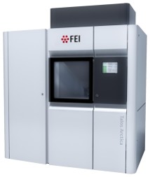 Model: Talos Arctica / Image courtesy: FEI Company |
|---|---|
| Microscopy | Model: FEI TALOS ARCTICA |
| Electron gun | Electron source:  FIELD EMISSION GUN / Accelerating voltage: 200 kV / Illumination mode: SPOT SCAN FIELD EMISSION GUN / Accelerating voltage: 200 kV / Illumination mode: SPOT SCAN |
| Electron lens | Mode: BRIGHT FIELD / Nominal magnification: 63000 X / Nominal defocus max: 3500 nm / Nominal defocus min: 1500 nm / Cs: 2.7 mm / C2 aperture diameter: 50 µm / Alignment procedure: COMA FREE |
| Specimen holder | Cryogen: NITROGEN |
| Image recording | Electron dose: 3.65 e/Å2 / Avg electron dose per subtomogram: 150 e/Å2 / Film or detector model: GATAN K3 BIOQUANTUM (6k x 4k) / Num. of real images: 1 |
| EM imaging optics | Energyfilter name: GIF Bioquantum / Details: Zero Loss Peak was refined after every tilt series / Energyfilter slit width: 20 eV |
- Processing
| EM software |
| ||||||||||||||||||||||||||||||||||||||||
|---|---|---|---|---|---|---|---|---|---|---|---|---|---|---|---|---|---|---|---|---|---|---|---|---|---|---|---|---|---|---|---|---|---|---|---|---|---|---|---|---|---|
| CTF correction | Type: PHASE FLIPPING AND AMPLITUDE CORRECTION | ||||||||||||||||||||||||||||||||||||||||
| Symmetry | Point symmetry: C2 (2 fold cyclic) | ||||||||||||||||||||||||||||||||||||||||
| 3D reconstruction | Resolution: 7 Å / Resolution method: FSC 0.143 CUT-OFF / Num. of particles: 9767 / Symmetry type: POINT | ||||||||||||||||||||||||||||||||||||||||
| EM volume selection | Method: Manually annotated / Num. of tomograms: 24 / Num. of volumes extracted: 340688 / Reference model: Ab-initio | ||||||||||||||||||||||||||||||||||||||||
| Atomic model building | B value: 351 / Protocol: RIGID BODY FIT / Space: REAL / Target criteria: CC Details: The initial model of 1 M Dimer was generated with AlphaFold2. Model was docked into map in Chimera v1.1.4. RealSpaceRefine in Phenix of 1 M Dimer. Open refined model and map in Chimera. ...Details: The initial model of 1 M Dimer was generated with AlphaFold2. Model was docked into map in Chimera v1.1.4. RealSpaceRefine in Phenix of 1 M Dimer. Open refined model and map in Chimera. Duplicate model, rotate and dock. Re run RealSpaceRefine. Repeat for all dimers present in electron density map. Run comprehensive validation in Phenix which resulted in Ramachandran outliers of 0%, Allowed 7% and Favored of 93% . We then removed all side chains from model prior to PDB deposition since our map does not support any side chain density - only backbone. | ||||||||||||||||||||||||||||||||||||||||
| Atomic model building | Source name: AlphaFold / Type: in silico model | ||||||||||||||||||||||||||||||||||||||||
| Refinement | Cross valid method: THROUGHOUT | ||||||||||||||||||||||||||||||||||||||||
| Displacement parameters | Biso max: 384.78 Å2 / Biso mean: 217.9891 Å2 / Biso min: 141.26 Å2 |
