 Movie
Movie Controller


+ Open data

- Basic information
| Entry | Database: PDB / ID: 9n01 | |||||||||
|---|---|---|---|---|---|---|---|---|---|---|
| Title | Cryo-EM structure of SeAvs7 MCP EFTu1 monomeric complex | |||||||||
 Components Components |
| |||||||||
 Keywords Keywords | ANTIVIRAL PROTEIN / AAA family ATPase | |||||||||
| Function / homology |  Function and homology information Function and homology informationguanyl-nucleotide exchange factor complex / protein-synthesizing GTPase / guanosine tetraphosphate binding / translational elongation / translation elongation factor activity / GTPase activity / GTP binding / magnesium ion binding / ATP hydrolysis activity / ATP binding ...guanyl-nucleotide exchange factor complex / protein-synthesizing GTPase / guanosine tetraphosphate binding / translational elongation / translation elongation factor activity / GTPase activity / GTP binding / magnesium ion binding / ATP hydrolysis activity / ATP binding / plasma membrane / cytoplasm Similarity search - Function | |||||||||
| Biological species |  Salmonella enterica (bacteria) Salmonella enterica (bacteria) Escherichia coli (E. coli) Escherichia coli (E. coli) Escherichia phage ZL19 (virus) Escherichia phage ZL19 (virus) | |||||||||
| Method | ELECTRON MICROSCOPY / single particle reconstruction / cryo EM / Resolution: 3.67 Å | |||||||||
 Authors Authors | Zhang, J. / Feng, L. | |||||||||
| Funding support | 1items
| |||||||||
 Citation Citation | Journal: Nature / Year: 2026 Title: Diverse bacterial pattern recognition receptors sense the core phage proteome. Authors: Hyunbin Lee / Sofia Luengo-Woods / Jianxiu Zhang / Kira S Makarova / Yuri I Wolf / Collin Chiu / Simone A Evans / Junyi Chen / Haopeng Xiao / Liang Feng / Eugene V Koonin / Alex Gao /  Abstract: Recognition of foreign molecules inside cells is critical for immunity across all domains of life. Proteins of the STAND NTPase superfamily, including eukaryotic NOD-like receptors, play a central ...Recognition of foreign molecules inside cells is critical for immunity across all domains of life. Proteins of the STAND NTPase superfamily, including eukaryotic NOD-like receptors, play a central role in this process. In bacteria and archaea, although several STAND families sense phage proteins, their functional diversity remains largely unexplored. Here we conduct a systematic phylogenetic analysis of prokaryotic STAND NTPases and identify at least 90 structurally distinct families associated with antiviral defence. We first show that the uncharacterized Avs7 family recognizes the major capsid protein (MCP) of tailed phages. Three cryogenic electron microscopy structures of Salmonella enterica Avs7 reveal an asymmetric, butterfly-shaped tetramer that assembles stepwise through large, MCP-induced conformational changes, incorporating bacterial elongation factor Tu (EF-Tu) as a structural component that enhances defence. Using genetic screens with a library of 687 phage genes, we further show that 13 additional STAND families sense 13 conserved phage proteins, encompassing most of the core structural and replicative components of tailed phages. These include 2 distinct MCP-sensing families (Avs8 and Avs10) and 11 others (Avs11-21), which recognize the portal, portal adaptor, tail nozzle, head-tail connector, tail terminator, tail tube protein, tail assembly chaperone, tape measure protein, DNA polymerase, helicase/RecA-type ATPase and single-stranded DNA annealing protein, respectively. Together, our findings reveal a mechanism of host-factor repurposing and establish structure-based pattern recognition as a fundamental strategy of bacterial immunity. | |||||||||
| History |
|
- Structure visualization
| Structure viewer | Molecule: MolmilJmol/JSmol |
|---|
- Downloads & links
-Download
| PDBx/mmCIF format | 9n01.cif.gz | 313.5 KB | Display | PDBx/mmCIF format |
|---|---|---|---|---|
| PDB format | pdb9n01.ent.gz | 241 KB | Display | PDB format |
| PDBx/mmJSON format | 9n01.json.gz | Tree view | PDBx/mmJSON format | |
| Others |  Other downloads Other downloads |
-Validation report
| Arichive directory | https://data.pdbj.org/pub/pdb/validation_reports/n0/9n01ftp://data.pdbj.org/pub/pdb/validation_reports/n0/9n01 | HTTPS FTP |
|---|
-Related structure data
| Related structure data |  48772MC  9n00C  9yixC M: map data used to model this data C: citing same article ( |
|---|---|
| Similar structure data |


-Links
 PDBj
PDBj

- Assembly
| Deposited unit | 
|
|---|---|
| 1 |
|
-Components
| #1: Protein | Mass: 172696.516 Da / Num. of mol.: 1 Source method: isolated from a genetically manipulated source Source: (gene. exp.) Salmonella enterica (bacteria) / Gene: DUC28_22665 / Production host: Escherichia coli BL21(DE3) (bacteria) / References: UniProt: A0A5H7DAD8 |
|---|---|
| #2: Protein | Mass: 43340.465 Da / Num. of mol.: 1 Source method: isolated from a genetically manipulated source Source: (gene. exp.) Escherichia coli (E. coli) / Gene: tufA, b3339, JW3301 / Production host: Escherichia coli BL21(DE3) (bacteria) / References: UniProt: P0CE47 |
| #3: Protein | Mass: 34455.023 Da / Num. of mol.: 1 Source method: isolated from a genetically manipulated source Source: (gene. exp.) Escherichia phage ZL19 (virus) / Production host: Escherichia coli BL21(DE3) (bacteria) / References: UniProt: A0A9E6YYI9 |
| #4: Chemical | ChemComp-GTP / 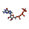 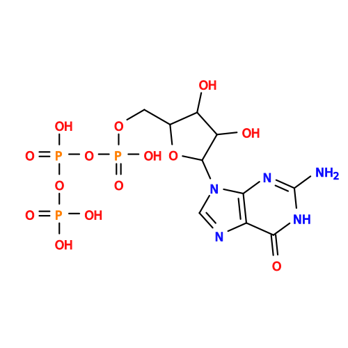 Mass: 523.180 Da / Num. of mol.: 1 / Source method: obtained synthetically / Formula: C10H16N5O14P3 / Feature type: SUBJECT OF INVESTIGATION / Comment: GTP, energy-carrying molecule*YM Mass: 523.180 Da / Num. of mol.: 1 / Source method: obtained synthetically / Formula: C10H16N5O14P3 / Feature type: SUBJECT OF INVESTIGATION / Comment: GTP, energy-carrying molecule*YM |
| Has ligand of interest | Y |
| Has protein modification | N |
-Experimental details
-Experiment
| Experiment | Method: ELECTRON MICROSCOPY |
|---|---|
| EM experiment | Aggregation state: PARTICLE / 3D reconstruction method: single particle reconstruction |
- Sample preparation
| Component | Name: Cryo-EM structure of SeAvs7 MCP EFTu1 monomeric complex Type: COMPLEX / Entity ID: #1, #3 / Source: RECOMBINANT | ||||||||||||
|---|---|---|---|---|---|---|---|---|---|---|---|---|---|
| Molecular weight | Experimental value: NO | ||||||||||||
| Source (natural) |
| ||||||||||||
| Source (recombinant) | Organism: Escherichia coli BL21(DE3) (bacteria) | ||||||||||||
| Buffer solution | pH: 7.4 | ||||||||||||
| Specimen | Embedding applied: NO / Shadowing applied: NO / Staining applied: NO / Vitrification applied: YES | ||||||||||||
| Vitrification | Cryogen name: ETHANE |
 Homo sapiens (human)
- Electron microscopy imaging
| Experimental equipment |  Model: Titan Krios / Image courtesy: FEI Company |
|---|---|
| Microscopy | Model: TFS KRIOS |
| Electron gun | Electron source:  FIELD EMISSION GUN / Accelerating voltage: 300 kV / Illumination mode: FLOOD BEAM FIELD EMISSION GUN / Accelerating voltage: 300 kV / Illumination mode: FLOOD BEAM |
| Electron lens | Mode: BRIGHT FIELD / Nominal defocus max: 2000 nm / Nominal defocus min: 1200 nm / Cs: 2.7 mm / C2 aperture diameter: 100 µm |
| Image recording | Electron dose: 50 e/Å2 / Film or detector model: FEI FALCON IV (4k x 4k) |
- Processing
| EM software |
| ||||||||||||||||||||||||
|---|---|---|---|---|---|---|---|---|---|---|---|---|---|---|---|---|---|---|---|---|---|---|---|---|---|
| CTF correction | Type: NONE | ||||||||||||||||||||||||
| 3D reconstruction | Resolution: 3.67 Å / Resolution method: FSC 0.143 CUT-OFF / Num. of particles: 106963 / Symmetry type: POINT | ||||||||||||||||||||||||
| Refinement | Stereochemistry target values: REAL-SPACE (WEIGHTED MAP SUM AT ATOM CENTERS) | ||||||||||||||||||||||||
| Refine LS restraints |
|
