 Movie
Movie Controller


+ Open data

- Basic information
| Entry | Database: PDB / ID: 9mqn | ||||||
|---|---|---|---|---|---|---|---|
| Title | AngV-F Pre-fusion Protein | ||||||
 Components Components | Fusion protein | ||||||
 Keywords Keywords | VIRAL PROTEIN / Henipavirus / AngV-F protein / Pre-fusion F protein / Cryo-EM structure / Fusion Ectodomain / Dimer-of-trimer | ||||||
| Biological species |  Angavokely henipavirus Angavokely henipavirus | ||||||
| Method | ELECTRON MICROSCOPY / single particle reconstruction / cryo EM / Resolution: 5.9 Å | ||||||
 Authors Authors | Lella, M. / Acharya, P. | ||||||
| Funding support |  United States, 1items United States, 1items
| ||||||
 Citation Citation | Journal: bioRxiv / Year: 2025 Title: Structural and antigenic characterization of novel and diverse Henipavirus glycoproteins. Abstract: Henipaviruses, a genus within the Paramyxoviridae family, include the highly virulent Nipah and Hendra viruses that cause reoccurring outbreaks of deadly disease. Recent discoveries of several new ...Henipaviruses, a genus within the Paramyxoviridae family, include the highly virulent Nipah and Hendra viruses that cause reoccurring outbreaks of deadly disease. Recent discoveries of several new Paramyxoviridae species, including the zoonotic Langya virus, have revealed much higher antigenic diversity than currently characterized and prompted the reorganization of these viruses into the Henipavirus and Parahenipavirus genera. Here, to explore the limits of structural and antigenic variation in both genera, collectively referred to here as HNVs, we constructed an expanded, antigenically diverse panel of HNV fusion and attachment glycoproteins from 56 unique HNV strains that better reflects global HNV diversity. We expressed and purified the fusion protein ectodomains and the attachment protein head domains and characterized their biochemical, biophysical and structural properties. We performed immunization experiments in mice leading to the elicitation of antibodies reactive to multiple HNV fusion proteins. Cryo-electron microscopy structures of diverse fusion proteins elucidated molecular determinants of differential pre-fusion state metastability and higher order contacts. A crystal structure of the Gamak virus attachment head domain revealed an additional domain added to the conserved 6-bladed, β-propeller fold. Taken together, these studies expand the known structural and antigenic limits of the HNVs, reveal new cross-reactive epitopes within both genera and provide foundational data for the development of broadly reactive countermeasures. | ||||||
| History |
|
- Structure visualization
| Structure viewer | Molecule:  MolmilJmol/JSmol MolmilJmol/JSmol |
|---|
- Downloads & links
-Download
| PDBx/mmCIF format | 9mqn.cif.gz | 473 KB | Display | PDBx/mmCIF format |
|---|---|---|---|---|
| PDB format | pdb9mqn.ent.gz | 394 KB | Display | PDB format |
| PDBx/mmJSON format | 9mqn.json.gz | Tree view | PDBx/mmJSON format | |
| Others |  Other downloads Other downloads |
-Validation report
| Arichive directory | https://data.pdbj.org/pub/pdb/validation_reports/mq/9mqnftp://data.pdbj.org/pub/pdb/validation_reports/mq/9mqn | HTTPS FTP |
|---|
-Related structure data
| Related structure data |  48535MC 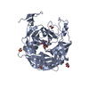 9ehuC 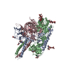 9mnhC M: map data used to model this data C: citing same article ( |
|---|
-Links
 PDBj
PDBj- Assembly
| Deposited unit | 
|
|---|---|
| 1 |
|
| Details | Dimer of trimer |
-Components
| #1: Protein | Mass: 58587.699 Da / Num. of mol.: 6 / Fragment: ectodomain Source method: isolated from a genetically manipulated source Details: the following domains are at at C-terminus: foldon trimerization domain, HRV3C protease site, His-tag, and two streptavidin tags separated by GS linker Source: (gene. exp.) Angavokely henipavirus / Production host:  Homo sapiens (human) Homo sapiens (human)#2: Polysaccharide | beta-D-mannopyranose-(1-4)-2-acetamido-2-deoxy-beta-D-glucopyranose-(1-4)-[alpha-L-fucopyranose-(1- ...beta-D-mannopyranose-(1-4)-2-acetamido-2-deoxy-beta-D-glucopyranose-(1-4)-[alpha-L-fucopyranose-(1-6)]2-acetamido-2-deoxy-beta-D-glucopyranose Source method: isolated from a genetically manipulated source #3: Sugar | ChemComp-NAG /   Type: D-saccharide, beta linking / Mass: 221.208 Da / Num. of mol.: 24 Type: D-saccharide, beta linking / Mass: 221.208 Da / Num. of mol.: 24Source method: isolated from a genetically manipulated source Formula: C8H15NO6 / Feature type: SUBJECT OF INVESTIGATION Compound details | pre-fusion conformation | Has ligand of interest | Y | Has protein modification | Y | |
|---|
-Experimental details
-Experiment
| Experiment | Method: ELECTRON MICROSCOPY |
|---|---|
| EM experiment | Aggregation state: 3D ARRAY / 3D reconstruction method: single particle reconstruction |
- Sample preparation
| Component | Name: Dimer-of-Trimer of AngV-F ectodomain in pre-fusion conformation Type: COMPLEX Details: AngV-F fusion ectodomain is expressed with following domains at C-terminus, foldon trimerization domain, HRV3C protease site, His-tag, and two streptavidin tags separated by GS linker Entity ID: #1 / Source: RECOMBINANT |
|---|---|
| Molecular weight | Value: 0.06 MDa / Experimental value: YES |
| Source (natural) | Organism: Angavokely henipavirus / Cellular location: Outer Membrane |
| Source (recombinant) | Organism: Homo sapiens (human) / Cell: HEK293 / Plasmid: palphaH |
| Buffer solution | pH: 7.4 |
| Buffer component | Conc.: 1 1 / Name: Phosphate Buffer / Formula: Na2HPO4/NaH2PO4 |
| Specimen | Conc.: 1.5 mg/ml / Embedding applied: NO / Shadowing applied: NO / Staining applied: NO / Vitrification applied: YES |
| Specimen support | Grid material: COPPER / Grid mesh size: 300 divisions/in. / Grid type: Quantifoil R1.2/1.3 |
| Vitrification | Instrument: LEICA EM GP / Cryogen name: ETHANE / Humidity: 95 % / Chamber temperature: 298 K Details: Protein sample was applied to the grid and incubated for 30 seconds at >95% humidity. Excess protein was blotted away for 2.5 seconds before being plunge frozen into liquid ethane |
- Electron microscopy imaging
| Experimental equipment |  Model: Titan Krios / Image courtesy: FEI Company |
|---|---|
| Microscopy | Model: TFS KRIOS / Details: Grid screening was performed manually |
| Electron gun | Electron source:  FIELD EMISSION GUN / Accelerating voltage: 300 kV / Illumination mode: FLOOD BEAM FIELD EMISSION GUN / Accelerating voltage: 300 kV / Illumination mode: FLOOD BEAM |
| Electron lens | Mode: BRIGHT FIELD / Nominal magnification: 81000 X / Calibrated magnification: 81000 X / Nominal defocus max: 2800 nm / Nominal defocus min: 1700 nm / Calibrated defocus min: 1700 nm / Calibrated defocus max: 2800 nm / Cs: 2.7 mm / C2 aperture diameter: 50 µm / Alignment procedure: COMA FREE |
| Specimen holder | Cryogen: NITROGEN / Specimen holder model: FEI TITAN KRIOS AUTOGRID HOLDER |
| Image recording | Average exposure time: 2.8 sec. / Electron dose: 28 e/Å2 / Film or detector model: GATAN K3 (6k x 4k) / Num. of grids imaged: 3 / Num. of real images: 24000 Details: Images were collected in a movie -mode at 22 frames per second |
| Image scans | Width: 5760 / Height: 4092 |
- Processing
| EM software |
| ||||||||||||||||||||||||||||
|---|---|---|---|---|---|---|---|---|---|---|---|---|---|---|---|---|---|---|---|---|---|---|---|---|---|---|---|---|---|
| Image processing | Details: Micrographs were motion-corrected and aligned micrographs were used for the processing | ||||||||||||||||||||||||||||
| CTF correction | Details: Micrographs were curated through contrast transfer function (CTF) where greater than 30 angstrom were discarded Type: NONE | ||||||||||||||||||||||||||||
| Particle selection | Num. of particles selected: 13247526 Details: Particles were picked using automated blob picker and extracted | ||||||||||||||||||||||||||||
| Symmetry | Point symmetry: C1 (asymmetric) | ||||||||||||||||||||||||||||
| 3D reconstruction | Resolution: 5.9 Å / Resolution method: FSC 0.143 CUT-OFF / Num. of particles: 240865 / Algorithm: FOURIER SPACE / Num. of class averages: 48 / Symmetry type: POINT | ||||||||||||||||||||||||||||
| Atomic model building |
| ||||||||||||||||||||||||||||
| Refinement | Highest resolution: 5.9 Å Stereochemistry target values: REAL-SPACE (WEIGHTED MAP SUM AT ATOM CENTERS) |
