[English] 日本語
 Yorodumi
- PDB-9lts: Cryo-EM structure of the Dinoroseobacter shibae RC-LH1 supercomplex -
+ Open data
- Basic information
| Entry | Database: PDB / ID: 9lts | |||||||||||||||||||||
|---|---|---|---|---|---|---|---|---|---|---|---|---|---|---|---|---|---|---|---|---|---|---|
| Title | Cryo-EM structure of the Dinoroseobacter shibae RC-LH1 supercomplex | |||||||||||||||||||||
 Components Components |
| |||||||||||||||||||||
 Keywords Keywords | PHOTOSYNTHESIS / reaction centre light-harvesting 1 | |||||||||||||||||||||
| Function / homology |  Function and homology information Function and homology informationorganelle inner membrane / plasma membrane-derived chromatophore membrane / plasma membrane light-harvesting complex / bacteriochlorophyll binding / photosynthetic electron transport in photosystem II / photosynthesis, light reaction / electron transfer activity / iron ion binding / heme binding / metal ion binding ...organelle inner membrane / plasma membrane-derived chromatophore membrane / plasma membrane light-harvesting complex / bacteriochlorophyll binding / photosynthetic electron transport in photosystem II / photosynthesis, light reaction / electron transfer activity / iron ion binding / heme binding / metal ion binding / membrane / plasma membrane Similarity search - Function | |||||||||||||||||||||
| Biological species |  Dinoroseobacter shibae DFL 12 = DSM 16493 (bacteria) Dinoroseobacter shibae DFL 12 = DSM 16493 (bacteria) | |||||||||||||||||||||
| Method | ELECTRON MICROSCOPY / single particle reconstruction / cryo EM / Resolution: 2.49 Å | |||||||||||||||||||||
 Authors Authors | Liu, Z.K. / Wang, P. / Liu, L.N. | |||||||||||||||||||||
| Funding support |  China, 1items China, 1items
| |||||||||||||||||||||
 Citation Citation |  Journal: To Be Published Journal: To Be PublishedTitle: Cryo-EM structure of the Dinoroseobacter shibae RC-LH1 supercomplex Authors: Liu, Z.K. / Wang, P. / Liu, L.N. | |||||||||||||||||||||
| History |
|
- Structure visualization
| Structure viewer | Molecule:  Molmil Molmil Jmol/JSmol Jmol/JSmol |
|---|
- Downloads & links
- Download
| PDBx/mmCIF format |  9lts.cif.gz 9lts.cif.gz | 693.3 KB | Display |  PDBx/mmCIF format PDBx/mmCIF format |
|---|---|---|---|---|
| PDB format |  pdb9lts.ent.gz pdb9lts.ent.gz | 596.3 KB | Display |  PDB format PDB format |
| PDBx/mmJSON format |  9lts.json.gz 9lts.json.gz | Tree view |  PDBx/mmJSON format PDBx/mmJSON format | |
| Others |  Other downloads Other downloads |
-Validation report
| Arichive directory |  https://data.pdbj.org/pub/pdb/validation_reports/lt/9lts https://data.pdbj.org/pub/pdb/validation_reports/lt/9lts ftp://data.pdbj.org/pub/pdb/validation_reports/lt/9lts ftp://data.pdbj.org/pub/pdb/validation_reports/lt/9lts | HTTPS FTP |
|---|
-Related structure data
| Related structure data |  63379MC M: map data used to model this data C: citing same article ( |
|---|---|
| Similar structure data | Similarity search - Function & homology  F&H Search F&H Search |
- Links
Links
- Assembly
| Deposited unit | 
|
|---|---|
| 1 |
|
- Components
-Protein , 2 types, 2 molecules AC
| #1: Protein | Mass: 7963.479 Da / Num. of mol.: 1 / Source method: isolated from a natural source Source: (natural)  Dinoroseobacter shibae DFL 12 = DSM 16493 (bacteria) Dinoroseobacter shibae DFL 12 = DSM 16493 (bacteria)References: UniProt: A8LIU2 |
|---|---|
| #4: Protein | Mass: 39580.883 Da / Num. of mol.: 1 / Source method: isolated from a natural source Source: (natural)  Dinoroseobacter shibae DFL 12 = DSM 16493 (bacteria) Dinoroseobacter shibae DFL 12 = DSM 16493 (bacteria)References: UniProt: A8LQ18 |
-Antenna pigment protein ... , 2 types, 34 molecules 153wusqomkgecai79264xvtrpnlhfd...
| #2: Protein | Mass: 5993.136 Da / Num. of mol.: 17 / Source method: isolated from a natural source Source: (natural)  Dinoroseobacter shibae DFL 12 = DSM 16493 (bacteria) Dinoroseobacter shibae DFL 12 = DSM 16493 (bacteria)References: UniProt: A8LQ15 #3: Protein/peptide | Mass: 5044.715 Da / Num. of mol.: 17 / Source method: isolated from a natural source Source: (natural)  Dinoroseobacter shibae DFL 12 = DSM 16493 (bacteria) Dinoroseobacter shibae DFL 12 = DSM 16493 (bacteria)References: UniProt: A8LQ14 |
|---|
-Reaction center protein ... , 3 types, 3 molecules HLM
| #5: Protein | Mass: 28621.299 Da / Num. of mol.: 1 / Source method: isolated from a natural source Source: (natural)  Dinoroseobacter shibae DFL 12 = DSM 16493 (bacteria) Dinoroseobacter shibae DFL 12 = DSM 16493 (bacteria)References: UniProt: A8LQ33 |
|---|---|
| #6: Protein | Mass: 30533.416 Da / Num. of mol.: 1 / Source method: isolated from a natural source Source: (natural)  Dinoroseobacter shibae DFL 12 = DSM 16493 (bacteria) Dinoroseobacter shibae DFL 12 = DSM 16493 (bacteria)References: UniProt: A8LQ16 |
| #7: Protein | Mass: 37037.172 Da / Num. of mol.: 1 / Source method: isolated from a natural source Source: (natural)  Dinoroseobacter shibae DFL 12 = DSM 16493 (bacteria) Dinoroseobacter shibae DFL 12 = DSM 16493 (bacteria)References: UniProt: A8LQ17 |
-Non-polymers , 7 types, 93 molecules 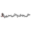
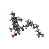
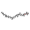
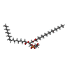

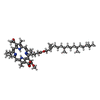


| #8: Chemical | ChemComp-U10 / #9: Chemical | ChemComp-BCL / #10: Chemical | ChemComp-SPN / #11: Chemical | ChemComp-MW9 / ( #12: Chemical | #13: Chemical | #14: Chemical | ChemComp-FE / | |
|---|
-Details
| Has ligand of interest | Y |
|---|---|
| Has protein modification | Y |
-Experimental details
-Experiment
| Experiment | Method: ELECTRON MICROSCOPY |
|---|---|
| EM experiment | Aggregation state: PARTICLE / 3D reconstruction method: single particle reconstruction |
- Sample preparation
| Component | Name: Cryo-EM structure of the Dinoroseobacter shibae RC-LH1 supercomplex Type: COMPLEX / Entity ID: #1-#7 / Source: NATURAL |
|---|---|
| Source (natural) | Organism:  Dinoroseobacter shibae DFL 12 = DSM 16493 (bacteria) Dinoroseobacter shibae DFL 12 = DSM 16493 (bacteria) |
| Buffer solution | pH: 8 |
| Specimen | Embedding applied: NO / Shadowing applied: NO / Staining applied: NO / Vitrification applied: YES |
| Vitrification | Cryogen name: ETHANE |
- Electron microscopy imaging
| Experimental equipment |  Model: Titan Krios / Image courtesy: FEI Company |
|---|---|
| Microscopy | Model: TFS KRIOS |
| Electron gun | Electron source:  FIELD EMISSION GUN / Accelerating voltage: 300 kV / Illumination mode: SPOT SCAN FIELD EMISSION GUN / Accelerating voltage: 300 kV / Illumination mode: SPOT SCAN |
| Electron lens | Mode: BRIGHT FIELD / Nominal defocus max: 1800 nm / Nominal defocus min: 800 nm |
| Image recording | Electron dose: 40 e/Å2 / Film or detector model: FEI FALCON IV (4k x 4k) |
- Processing
| EM software |
| |||||||||||||||||||||||||||||||||||||||||||||||||||||||||||||||
|---|---|---|---|---|---|---|---|---|---|---|---|---|---|---|---|---|---|---|---|---|---|---|---|---|---|---|---|---|---|---|---|---|---|---|---|---|---|---|---|---|---|---|---|---|---|---|---|---|---|---|---|---|---|---|---|---|---|---|---|---|---|---|---|---|
| Image processing |
| |||||||||||||||||||||||||||||||||||||||||||||||||||||||||||||||
| CTF correction |
| |||||||||||||||||||||||||||||||||||||||||||||||||||||||||||||||
| 3D reconstruction |
| |||||||||||||||||||||||||||||||||||||||||||||||||||||||||||||||
| Refinement | Highest resolution: 2.49 Å |
 Movie
Movie Controller


 PDBj
PDBj


