[English] 日本語
 Yorodumi
- PDB-9h4i: Cryo-EM structure of an octameric G10-resistosome from wheat (N-t... -
+ Open data
- Basic information
| Entry | Database: PDB / ID: 9h4i | ||||||
|---|---|---|---|---|---|---|---|
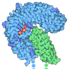
| Title | Cryo-EM structure of an octameric G10-resistosome from wheat (N-to-N arrangement) | ||||||
 Components Components | NB-ARC domain-containing protein | ||||||
 Keywords Keywords | PLANT PROTEIN / G10-NLR / Octamer / Resistance / LRR | ||||||
| Function / homology |  Function and homology information Function and homology informationinnate immune response-activating signaling pathway / plant-type hypersensitive response / ADP binding / defense response to bacterium / ATP binding Similarity search - Function | ||||||
| Biological species |  | ||||||
| Method | ELECTRON MICROSCOPY / single particle reconstruction / cryo EM / Resolution: 4.6 Å | ||||||
 Authors Authors | Guo, G.H. / Zhao, H. / Lukoyanova, N. / Selvaraj, M. / Jones, J. | ||||||
| Funding support |  United Kingdom, 1items United Kingdom, 1items
| ||||||
 Citation Citation |  Journal: Biorxiv / Year: 2025 Journal: Biorxiv / Year: 2025Title: An activated wheat CCG10-NLR immune receptor forms an octameric resistosome Authors: Guo, G. / Zhao, H. / Bai, K. / Wu, Q. / Dong, L. / Lu, L. / Chen, Y. / Hou, Y. / Lu, J. / Lu, P. / Li, M. / Zhang, H. / Wang, G. / Zhu, K. / Huang, B. / Cui, X. / Fu, H. / Hu, C. / Chu, Z. / ...Authors: Guo, G. / Zhao, H. / Bai, K. / Wu, Q. / Dong, L. / Lu, L. / Chen, Y. / Hou, Y. / Lu, J. / Lu, P. / Li, M. / Zhang, H. / Wang, G. / Zhu, K. / Huang, B. / Cui, X. / Fu, H. / Hu, C. / Chu, Z. / Lyu, X. / Kamoun, S. / Wang, C. / Liu, Z. / Selvaraj, M. / Jones, J.D.G. | ||||||
| History |
|
- Structure visualization
| Structure viewer | Molecule:  Molmil Molmil Jmol/JSmol Jmol/JSmol |
|---|
- Downloads & links
- Download
| PDBx/mmCIF format |  9h4i.cif.gz 9h4i.cif.gz | 2.3 MB | Display |  PDBx/mmCIF format PDBx/mmCIF format |
|---|---|---|---|---|
| PDB format |  pdb9h4i.ent.gz pdb9h4i.ent.gz | Display |  PDB format PDB format | |
| PDBx/mmJSON format |  9h4i.json.gz 9h4i.json.gz | Tree view |  PDBx/mmJSON format PDBx/mmJSON format | |
| Others |  Other downloads Other downloads |
-Validation report
| Arichive directory |  https://data.pdbj.org/pub/pdb/validation_reports/h4/9h4i https://data.pdbj.org/pub/pdb/validation_reports/h4/9h4i ftp://data.pdbj.org/pub/pdb/validation_reports/h4/9h4i ftp://data.pdbj.org/pub/pdb/validation_reports/h4/9h4i | HTTPS FTP |
|---|
-Related structure data
| Related structure data |  51852MC  9h2lC  9h73C M: map data used to model this data C: citing same article ( |
|---|---|
| Similar structure data | Similarity search - Function & homology  F&H Search F&H Search |
- Links
Links
- Assembly
| Deposited unit | 
|
|---|---|
| 1 |
|
- Components
| #1: Protein | Mass: 107700.422 Da / Num. of mol.: 16 Source method: isolated from a genetically manipulated source Source: (gene. exp.)   #2: Chemical | ChemComp-ATP / Has ligand of interest | Y | Has protein modification | N | |
|---|
-Experimental details
-Experiment
| Experiment | Method: ELECTRON MICROSCOPY |
|---|---|
| EM experiment | Aggregation state: PARTICLE / 3D reconstruction method: single particle reconstruction |
- Sample preparation
| Component | Name: Wheat auto immunity -3 Protein / Type: COMPLEX Details: This is a resistance protein in the wheat plant in an N-to-N arrangement, where N-terminal domains connect two octamers Entity ID: #1 / Source: RECOMBINANT |
|---|---|
| Molecular weight | Value: 1.712 MDa / Experimental value: NO |
| Source (natural) | Organism:  |
| Source (recombinant) | Organism:  |
| Buffer solution | pH: 7.5 Details: 150mM Tris HCl pH 7.5 150mM NaCl 1mM EDTA 1mM MgCl2 |
| Buffer component | Conc.: 150 mM / Name: Sodium chloride / Formula: NaCl |
| Specimen | Embedding applied: NO / Shadowing applied: NO / Staining applied: NO / Vitrification applied: YES Details: this is a protein sample purified from plant cell extract after over expression in leaf tissues |
| Specimen support | Grid type: Quantifoil R1.2/1.3 |
| Vitrification | Instrument: FEI VITROBOT MARK IV / Cryogen name: ETHANE / Humidity: 95 % / Chamber temperature: 277 K Details: 3 microlitre of the sample was applied on a graphene coated Quantafoil grid R1.2/1.3 copper grid and vitrification was performed using vitrobot |
- Electron microscopy imaging
| Experimental equipment |  Model: Titan Krios / Image courtesy: FEI Company |
|---|---|
| Microscopy | Model: TFS KRIOS |
| Electron gun | Electron source:  FIELD EMISSION GUN / Accelerating voltage: 300 kV / Illumination mode: FLOOD BEAM FIELD EMISSION GUN / Accelerating voltage: 300 kV / Illumination mode: FLOOD BEAM |
| Electron lens | Mode: BRIGHT FIELD / Nominal magnification: 105000 X / Nominal defocus max: 2500 nm / Nominal defocus min: 1300 nm / Cs: 2.7 mm / C2 aperture diameter: 50 µm / Alignment procedure: COMA FREE |
| Specimen holder | Cryogen: NITROGEN / Specimen holder model: FEI TITAN KRIOS AUTOGRID HOLDER |
| Image recording | Average exposure time: 1.94 sec. / Electron dose: 50 e/Å2 / Film or detector model: GATAN K3 (6k x 4k) / Num. of grids imaged: 1 / Num. of real images: 7044 Details: One grid was imaged at areas with good ice and several images |
| EM imaging optics | Energyfilter name: GIF Bioquantum / Energyfilter slit width: 10 eV |
- Processing
| EM software |
| ||||||||||||||||||||||||
|---|---|---|---|---|---|---|---|---|---|---|---|---|---|---|---|---|---|---|---|---|---|---|---|---|---|
| CTF correction | Type: PHASE FLIPPING ONLY | ||||||||||||||||||||||||
| Symmetry | Point symmetry: D8 (2x8 fold dihedral) | ||||||||||||||||||||||||
| 3D reconstruction | Resolution: 4.6 Å / Resolution method: FSC 0.143 CUT-OFF / Num. of particles: 13948 / Symmetry type: POINT | ||||||||||||||||||||||||
| Atomic model building | Protocol: FLEXIBLE FIT / Space: REAL Details: The atomic model was fitted flexibly using coot and refined using Phenix realspace refinement module | ||||||||||||||||||||||||
| Atomic model building | Source name: AlphaFold / Type: in silico model | ||||||||||||||||||||||||
| Refine LS restraints |
|
 Movie
Movie Controller


 PDBj
PDBj


