 Movie
Movie Controller


+ Open data

- Basic information
| Entry | Database: PDB / ID: 9fef | |||||||||||||||||||||||||||||||||
|---|---|---|---|---|---|---|---|---|---|---|---|---|---|---|---|---|---|---|---|---|---|---|---|---|---|---|---|---|---|---|---|---|---|---|
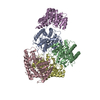
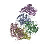
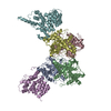
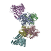
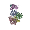
| Title | Cryo-EM structure of Trypanosoma cruzi (MDH)4-PEX5 complex | |||||||||||||||||||||||||||||||||
 Components Components |
| |||||||||||||||||||||||||||||||||
 Keywords Keywords | TRANSPORT PROTEIN / Peroxisomal protein transport / Cargo-Receptor complex | |||||||||||||||||||||||||||||||||
| Function / homology |  Function and homology information Function and homology informationintracellular organelle lumen / peroxisome matrix targeting signal-1 binding / protein import into peroxisome matrix, docking / (S)-malate dehydrogenase (NAD+, oxaloacetate-forming) / L-malate dehydrogenase (NAD+) activity / carboxylic acid metabolic process / peroxisomal membrane / tricarboxylic acid cycle / cytosol / cytoplasm Similarity search - Function | |||||||||||||||||||||||||||||||||
| Biological species |  | |||||||||||||||||||||||||||||||||
| Method | ELECTRON MICROSCOPY / single particle reconstruction / cryo EM / Resolution: 2.98 Å | |||||||||||||||||||||||||||||||||
 Authors Authors | Lipinski, O. / Sonani, R.R. / Blat, A. / Jemiola-Rzeminska, M. / Patel, S.N. / Sood, T. / Dubin, G. | |||||||||||||||||||||||||||||||||
| Funding support |  Poland, 3items Poland, 3items
| |||||||||||||||||||||||||||||||||
 Citation Citation | Journal: Nat Commun / Year: 2025 Title: Structure of Trypanosoma peroxisomal import complex unveils conformational heterogeneity. Authors: Ravi R Sonani / Artur Blat / Malgorzata Jemiola-Rzeminska / Oskar Lipinski / Stuti N Patel / Tabassum Sood / Grzegorz Dubin /   Abstract: Peroxisomes are membrane enclosed organelles hosting diverse metabolic processes in eukaryotic cells. Having no protein synthetic abilities, peroxisomes import all required enzymes from the cytosol ...Peroxisomes are membrane enclosed organelles hosting diverse metabolic processes in eukaryotic cells. Having no protein synthetic abilities, peroxisomes import all required enzymes from the cytosol through a peroxin (Pex) import system. Peroxisome targeting sequence 1 (PTS1)-tagged cargo is recognized by cytosolic receptor, Pex5. The cargo-Pex5 complex docks at the peroxisomal membrane translocon, composed of Pex14 and Pex13, facilitating translocation into the peroxisomal lumen. Despite its significance, the structural basis of the process is only partially understood. Here, we characterize the cargo-Pex5-Pex14 ternary complex from Trypanosoma cruzi. Cryo-electron microscopy maps enabled model building for Pex5 (residues 327-462 and 487-653) bound to malate dehydrogenase (MDH; residues 1-323) cargo tetramer and Pex14 (residues 21-85). The model provides insight into conformational heterogeneity and identifies secondary interfaces. Specifically, we observe that orientations of Pex5 relative to MDH span a 17° angle. Additionally, PTS1- and Wxxx(F/Y)-independent contact surfaces are observed at MDH-Pex5 and Pex5-Pex14 interfaces, respectively. Mutational analysis indicates that the non-PTS1 MDH-Pex5 interface does not significantly contribute to the affinity, but limits the conformational heterogeneity of MDH-Pex5 interface. The Pex5-Pex14 interface constitutes an extended binding site of Pex14 over Pex5. We discuss the implications of these findings for understanding peroxisomal import mechanism. #1: Journal: Biorxiv / Year: 2024Title: Structure of Trypanosoma peroxisomal import complex unveils conformational dynamics Authors: Sonani, R.R. / Blat, A. / Jemiola-Rzeminska, M. / Lipinski, O. / Patel, S.N. / Sood, T. / Dubin, G. | |||||||||||||||||||||||||||||||||
| History |
|
- Structure visualization
| Structure viewer | Molecule: MolmilJmol/JSmol |
|---|
- Downloads & links
-Download
| PDBx/mmCIF format | 9fef.cif.gz | 284.9 KB | Display | PDBx/mmCIF format |
|---|---|---|---|---|
| PDB format | pdb9fef.ent.gz | 225 KB | Display | PDB format |
| PDBx/mmJSON format | 9fef.json.gz | Tree view | PDBx/mmJSON format | |
| Others |  Other downloads Other downloads |
-Validation report
| Arichive directory | https://data.pdbj.org/pub/pdb/validation_reports/fe/9fefftp://data.pdbj.org/pub/pdb/validation_reports/fe/9fef | HTTPS FTP |
|---|
-Related structure data
| Related structure data | 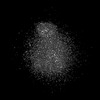 50340MC  9feeC C: citing same article ( M: map data used to model this data |
|---|---|
| Similar structure data |

-Links
 PDBj
PDBj


- Assembly
| Deposited unit | 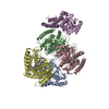
|
|---|---|
| 1 |
|
-Components
| #1: Protein | Mass: 35137.980 Da / Num. of mol.: 4 Source method: isolated from a genetically manipulated source Source: (gene. exp.) Gene: Tc00.1047053506503.69 / Production host:  References: UniProt: Q4DRD8, (S)-malate dehydrogenase (NAD+, oxaloacetate-forming) #2: Protein | | Mass: 75299.586 Da / Num. of mol.: 1 Source method: isolated from a genetically manipulated source Source: (gene. exp.) Gene: TCDM_08436 / Production host: Has protein modification | N | |
|---|
-Experimental details
-Experiment
| Experiment | Method: ELECTRON MICROSCOPY |
|---|---|
| EM experiment | Aggregation state: PARTICLE / 3D reconstruction method: single particle reconstruction |
- Sample preparation
| Component | Name: Ternary complex of (MDH)4 with PEX5 / Type: COMPLEX / Entity ID: all / Source: RECOMBINANT |
|---|---|
| Molecular weight | Value: 0.231 MDa / Experimental value: NO |
| Source (natural) | Organism: |
| Source (recombinant) | Organism: |
| Buffer solution | pH: 8 |
| Specimen | Embedding applied: NO / Shadowing applied: NO / Staining applied: NO / Vitrification applied: YES |
| Vitrification | Cryogen name: ETHANE |
- Electron microscopy imaging
| Experimental equipment |  Model: Titan Krios / Image courtesy: FEI Company |
|---|---|
| Microscopy | Model: FEI TITAN KRIOS |
| Electron gun | Electron source:  FIELD EMISSION GUN / Accelerating voltage: 300 kV / Illumination mode: FLOOD BEAM FIELD EMISSION GUN / Accelerating voltage: 300 kV / Illumination mode: FLOOD BEAM |
| Electron lens | Mode: BRIGHT FIELD / Nominal defocus max: 3380 nm / Nominal defocus min: 900 nm |
| Image recording | Electron dose: 42.79 e/Å2 / Film or detector model: GATAN K3 BIOQUANTUM (6k x 4k) |
- Processing
| EM software | Name: PHENIX / Version: 1.20.1_4487: / Category: model refinement | ||||||||||||||||||||||||
|---|---|---|---|---|---|---|---|---|---|---|---|---|---|---|---|---|---|---|---|---|---|---|---|---|---|
| CTF correction | Type: PHASE FLIPPING AND AMPLITUDE CORRECTION | ||||||||||||||||||||||||
| 3D reconstruction | Resolution: 2.98 Å / Resolution method: FSC 0.143 CUT-OFF / Num. of particles: 538505 / Symmetry type: POINT | ||||||||||||||||||||||||
| Refine LS restraints |
|
