[English] 日本語
 Yorodumi
- PDB-9cgi: Cryo-EM structure of the Nipah Virus polymerase (L) protein in co... -
+ Open data
- Basic information
| Entry | Database: PDB / ID: 9cgi | ||||||
|---|---|---|---|---|---|---|---|
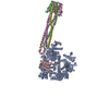
| Title | Cryo-EM structure of the Nipah Virus polymerase (L) protein in complex with the tetrameric phosphoprotein (P) | ||||||
 Components Components |
| ||||||
 Keywords Keywords | TRANSFERASE / VIRAL PROTEIN / Nipah virus / L protein / phosphoprotein / RNA-dependent RNA polymerase / PRNTase / GDP polyribonucleotidyl transferase / RNA capping / viral replication | ||||||
| Function / homology |  Function and homology information Function and homology informationnegative stranded viral RNA transcription / NNS virus cap methyltransferase / GDP polyribonucleotidyltransferase / negative stranded viral RNA replication / Hydrolases; Acting on acid anhydrides; In phosphorus-containing anhydrides / virion component / molecular adaptor activity / mRNA 5'-cap (guanine-N7-)-methyltransferase activity / host cell cytoplasm / symbiont-mediated suppression of host innate immune response ...negative stranded viral RNA transcription / NNS virus cap methyltransferase / GDP polyribonucleotidyltransferase / negative stranded viral RNA replication / Hydrolases; Acting on acid anhydrides; In phosphorus-containing anhydrides / virion component / molecular adaptor activity / mRNA 5'-cap (guanine-N7-)-methyltransferase activity / host cell cytoplasm / symbiont-mediated suppression of host innate immune response / RNA-directed RNA polymerase / RNA-directed RNA polymerase activity / GTPase activity / ATP binding Similarity search - Function | ||||||
| Biological species |  Henipavirus nipahense Henipavirus nipahense | ||||||
| Method | ELECTRON MICROSCOPY / single particle reconstruction / cryo EM / Resolution: 2.92 Å | ||||||
 Authors Authors | Liu, B. / Yang, G. / Wang, D. | ||||||
| Funding support | 1items
| ||||||
 Citation Citation |  Journal: Nat Commun / Year: 2024 Journal: Nat Commun / Year: 2024Title: Structure of the Nipah virus polymerase phosphoprotein complex. Authors: Ge Yang / Dong Wang / Bin Liu /  Abstract: The Nipah virus (NiV), a member of the Paramyxoviridae family, is notorious for its high fatality rate in humans. The RNA polymerase machinery of NiV, comprising the large protein L and the ...The Nipah virus (NiV), a member of the Paramyxoviridae family, is notorious for its high fatality rate in humans. The RNA polymerase machinery of NiV, comprising the large protein L and the phosphoprotein P, is essential for viral replication. This study presents the 2.9-Å cryo-electron microscopy structure of the NiV L-P complex, shedding light on its assembly and functionality. The structure not only demonstrates the molecular details of the conserved N-terminal domain, RNA-dependent RNA polymerase (RdRp), and GDP polyribonucleotidyltransferase of the L protein, but also the intact central oligomerization domain and the C-terminal X domain of the P protein. The P protein interacts extensively with the L protein, forming an antiparallel β-sheet among the P protomers and with the fingers subdomain of RdRp. The flexible linker domain of one P promoter extends its contact with the fingers subdomain to reach near the nascent RNA exit, highlighting the distinct characteristic of the NiV L-P interface. This distinctive tetrameric organization of the P protein and its interaction with the L protein provide crucial molecular insights into the replication and transcription mechanisms of NiV polymerase, ultimately contributing to the development of effective treatments and preventive measures against this Paramyxoviridae family deadly pathogen. | ||||||
| History |
|
- Structure visualization
| Structure viewer | Molecule:  Molmil Molmil Jmol/JSmol Jmol/JSmol |
|---|
- Downloads & links
- Download
| PDBx/mmCIF format |  9cgi.cif.gz 9cgi.cif.gz | 422.3 KB | Display |  PDBx/mmCIF format PDBx/mmCIF format |
|---|---|---|---|---|
| PDB format |  pdb9cgi.ent.gz pdb9cgi.ent.gz | 311.5 KB | Display |  PDB format PDB format |
| PDBx/mmJSON format |  9cgi.json.gz 9cgi.json.gz | Tree view |  PDBx/mmJSON format PDBx/mmJSON format | |
| Others |  Other downloads Other downloads |
-Validation report
| Summary document |  9cgi_validation.pdf.gz 9cgi_validation.pdf.gz | 1.2 MB | Display |  wwPDB validaton report wwPDB validaton report |
|---|---|---|---|---|
| Full document |  9cgi_full_validation.pdf.gz 9cgi_full_validation.pdf.gz | 1.2 MB | Display | |
| Data in XML |  9cgi_validation.xml.gz 9cgi_validation.xml.gz | 62.4 KB | Display | |
| Data in CIF |  9cgi_validation.cif.gz 9cgi_validation.cif.gz | 93.9 KB | Display | |
| Arichive directory |  https://data.pdbj.org/pub/pdb/validation_reports/cg/9cgi https://data.pdbj.org/pub/pdb/validation_reports/cg/9cgi ftp://data.pdbj.org/pub/pdb/validation_reports/cg/9cgi ftp://data.pdbj.org/pub/pdb/validation_reports/cg/9cgi | HTTPS FTP |
-Related structure data
| Related structure data |  45580MC M: map data used to model this data C: citing same article ( |
|---|---|
| Similar structure data | Similarity search - Function & homology  F&H Search F&H Search |
- Links
Links
- Assembly
| Deposited unit | 
|
|---|---|
| 1 |
|
- Components
| #1: Protein | Mass: 257565.156 Da / Num. of mol.: 1 Source method: isolated from a genetically manipulated source Source: (gene. exp.)  Henipavirus nipahense / Cell line (production host): Sf9 / Production host: Henipavirus nipahense / Cell line (production host): Sf9 / Production host:  References: UniProt: Q997F0, RNA-directed RNA polymerase, Hydrolases; Acting on acid anhydrides; In phosphorus-containing anhydrides, GDP polyribonucleotidyltransferase, NNS virus cap methyltransferase | ||
|---|---|---|---|
| #2: Protein | Mass: 78390.320 Da / Num. of mol.: 4 Source method: isolated from a genetically manipulated source Source: (gene. exp.)  Henipavirus nipahense / Gene: P/V/C / Cell line (production host): Sf9 / Production host: Henipavirus nipahense / Gene: P/V/C / Cell line (production host): Sf9 / Production host:  Has protein modification | Y | |
-Experimental details
-Experiment
| Experiment | Method: ELECTRON MICROSCOPY |
|---|---|
| EM experiment | Aggregation state: PARTICLE / 3D reconstruction method: single particle reconstruction |
- Sample preparation
| Component | Name: The Nipah virus L-P complex / Type: COMPLEX / Entity ID: all / Source: RECOMBINANT |
|---|---|
| Molecular weight | Value: 0.65 MDa / Experimental value: YES |
| Source (natural) | Organism:  Henipavirus nipahense Henipavirus nipahense |
| Source (recombinant) | Organism:  |
| Buffer solution | pH: 8 Details: 50 mM Tris-HCl pH 8.0, 250 mM NaCl, 5% glycerol, 1 mM TCEP, and 4 mM MgCl2 |
| Specimen | Embedding applied: NO / Shadowing applied: NO / Staining applied: NO / Vitrification applied: YES |
| Specimen support | Grid material: COPPER / Grid mesh size: 300 divisions/in. / Grid type: Quantifoil R1.2/1.3 |
| Vitrification | Instrument: FEI VITROBOT MARK IV / Cryogen name: ETHANE / Humidity: 100 % / Chamber temperature: 277 K |
- Electron microscopy imaging
| Experimental equipment |  Model: Titan Krios / Image courtesy: FEI Company |
|---|---|
| Microscopy | Model: FEI TITAN KRIOS |
| Electron gun | Electron source:  FIELD EMISSION GUN / Accelerating voltage: 300 kV / Illumination mode: FLOOD BEAM FIELD EMISSION GUN / Accelerating voltage: 300 kV / Illumination mode: FLOOD BEAM |
| Electron lens | Mode: BRIGHT FIELD / Nominal magnification: 130000 X / Nominal defocus max: 2000 nm / Nominal defocus min: 1000 nm / Cs: 2.7 mm / C2 aperture diameter: 50 µm / Alignment procedure: COMA FREE |
| Specimen holder | Cryogen: NITROGEN / Specimen holder model: FEI TITAN KRIOS AUTOGRID HOLDER / Temperature (max): 77 K / Temperature (min): 63 K |
| Image recording | Average exposure time: 1.7 sec. / Electron dose: 54.8 e/Å2 / Film or detector model: GATAN K3 BIOQUANTUM (6k x 4k) / Num. of grids imaged: 1 / Num. of real images: 6114 |
| Image scans | Width: 5760 / Height: 4092 |
- Processing
| EM software | Name: PHENIX / Version: 1.21_5207: / Category: model refinement | ||||||||||||||||||||||||
|---|---|---|---|---|---|---|---|---|---|---|---|---|---|---|---|---|---|---|---|---|---|---|---|---|---|
| CTF correction | Type: PHASE FLIPPING AND AMPLITUDE CORRECTION | ||||||||||||||||||||||||
| Particle selection | Num. of particles selected: 3052781 | ||||||||||||||||||||||||
| Symmetry | Point symmetry: C1 (asymmetric) | ||||||||||||||||||||||||
| 3D reconstruction | Resolution: 2.92 Å / Resolution method: FSC 0.143 CUT-OFF / Num. of particles: 278295 / Algorithm: SIMULTANEOUS ITERATIVE (SIRT) / Num. of class averages: 1 / Symmetry type: POINT | ||||||||||||||||||||||||
| Atomic model building | B value: 73.5 / Protocol: OTHER / Space: REAL | ||||||||||||||||||||||||
| Refine LS restraints |
|
 Movie
Movie Controller


 PDBj
PDBj


