 Movie
Movie Controller

[English] 日本語
 Yorodumi
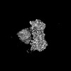
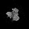
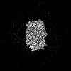
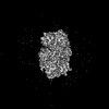
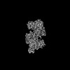
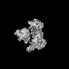
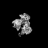
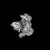
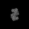
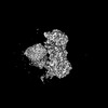
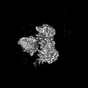
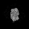
- PDB-9byh: Consensus model for turnover condition of Bacillus subtilis ribon... -
+ Open data

- Basic information
| Entry | Database: PDB / ID: 9byh | |||||||||
|---|---|---|---|---|---|---|---|---|---|---|
| Title | Consensus model for turnover condition of Bacillus subtilis ribonucleotide reductase complex | |||||||||
 Components Components | (Ribonucleoside-diphosphate reductase subunit ...) x 2 | |||||||||
 Keywords Keywords | OXIDOREDUCTASE / Ribonucleotide Reductase | |||||||||
| Function / homology |  Function and homology information Function and homology informationribonucleoside-diphosphate reductase complex / ribonucleoside-diphosphate reductase / ribonucleoside-diphosphate reductase activity, thioredoxin disulfide as acceptor / deoxyribonucleotide biosynthetic process / ATP binding / metal ion binding Similarity search - Function | |||||||||
| Biological species |  | |||||||||
| Method | ELECTRON MICROSCOPY / single particle reconstruction / cryo EM / Resolution: 2.53 Å | |||||||||
 Authors Authors | Xu, D. / Thomas, W.C. / Burnim, A.A. / Ando, N. | |||||||||
| Funding support |  United States, 2items United States, 2items
| |||||||||
 Citation Citation | Journal: Nat Commun / Year: 2025 Title: Conformational landscapes of a class I ribonucleotide reductase complex during turnover reveal intrinsic dynamics and asymmetry. Authors: Da Xu / William C Thomas / Audrey A Burnim / Nozomi Ando / Abstract: Understanding the structural dynamics associated with enzymatic catalysis has been a long-standing goal of biochemistry. With the advent of modern cryo-electron microscopy (cryo-EM), it has become ...Understanding the structural dynamics associated with enzymatic catalysis has been a long-standing goal of biochemistry. With the advent of modern cryo-electron microscopy (cryo-EM), it has become conceivable to redefine a protein's structure as the continuum of all conformations and their distributions. However, capturing and interpreting this information remains challenging. Here, we use classification and deep-learning-based analyses to characterize the conformational heterogeneity of a class I ribonucleotide reductase (RNR) during turnover. By converting the resulting information into physically interpretable 2D conformational landscapes, we demonstrate that RNR continuously samples a wide range of motions while maintaining surprising asymmetry to regulate the two halves of its turnover cycle. Remarkably, we directly observe the appearance of highly transient conformations needed for catalysis, as well as the interaction of RNR with its endogenous reductant thioredoxin also contributing to the asymmetry and dynamics of the enzyme complex. Overall, this work highlights the role of conformational dynamics in regulating key steps in enzyme mechanisms. | |||||||||
| History |
|
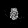
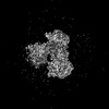
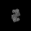
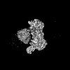
- Structure visualization
| Structure viewer | Molecule: MolmilJmol/JSmol |
|---|
- Downloads & links
-Download
| PDBx/mmCIF format | 9byh.cif.gz | 300.1 KB | Display | PDBx/mmCIF format |
|---|---|---|---|---|
| PDB format | pdb9byh.ent.gz | 234.6 KB | Display | PDB format |
| PDBx/mmJSON format | 9byh.json.gz | Tree view | PDBx/mmJSON format | |
| Others |  Other downloads Other downloads |
-Validation report
| Arichive directory | https://data.pdbj.org/pub/pdb/validation_reports/by/9byhftp://data.pdbj.org/pub/pdb/validation_reports/by/9byh | HTTPS FTP |
|---|
-Related structure data
| Related structure data |  45031MC  9bw3C  9bwxC  9bx2C  9bx3C  9bx6C  9bx8C  9bx9C  9bxcC  9bxsC  9bxtC  9bxxC  9bxzC  9by0C  9by1C  9by2C  9by3C  9by7C  9by8C  9by9C  9byaC  9bycC 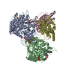 9bydC 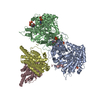 9bygC 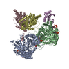 9bylC  9bytC  9byvC  9bywC  9byxC  9byyC  9byzC  9bz2C  9bz3C  9bz5C  9bz6C  9bz9C  9bzaC  9bzdC 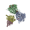 9bzeC 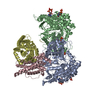 9bzfC  9bzhC  9bziC  9bzjC 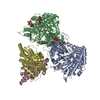 9bzkC  9bzmC  9bzoC M: map data used to model this data C: citing same article ( |
|---|---|
| Similar structure data |


-Links
 PDBj
PDBj
- Assembly
| Deposited unit | 
|
|---|---|
| 1 |
|
-Components
-Ribonucleoside-diphosphate reductase subunit ... , 2 types, 4 molecules ABCD
| #1: Protein | Mass: 80791.469 Da / Num. of mol.: 2 Source method: isolated from a genetically manipulated source Source: (gene. exp.) References: UniProt: P50620, ribonucleoside-diphosphate reductase #2: Protein | Mass: 40720.992 Da / Num. of mol.: 2 Source method: isolated from a genetically manipulated source Source: (gene. exp.) References: UniProt: P50621, ribonucleoside-diphosphate reductase |
|---|
-Non-polymers , 4 types, 8 molecules 


| #3: Chemical |  Mass: 507.181 Da / Num. of mol.: 2 / Source method: obtained synthetically / Formula: C10H16N5O13P3 / Feature type: SUBJECT OF INVESTIGATION / Comment: ATP, energy-carrying molecule*YM Mass: 507.181 Da / Num. of mol.: 2 / Source method: obtained synthetically / Formula: C10H16N5O13P3 / Feature type: SUBJECT OF INVESTIGATION / Comment: ATP, energy-carrying molecule*YM#4: Chemical |  Type: RNA linking / Mass: 443.201 Da / Num. of mol.: 2 / Source method: obtained synthetically / Formula: C10H15N5O11P2 / Feature type: SUBJECT OF INVESTIGATION / Comment: GDP, energy-carrying molecule*YM Type: RNA linking / Mass: 443.201 Da / Num. of mol.: 2 / Source method: obtained synthetically / Formula: C10H15N5O11P2 / Feature type: SUBJECT OF INVESTIGATION / Comment: GDP, energy-carrying molecule*YM#5: Chemical |  Mass: 482.168 Da / Num. of mol.: 2 / Source method: obtained synthetically / Formula: C10H17N2O14P3 / Feature type: SUBJECT OF INVESTIGATION Mass: 482.168 Da / Num. of mol.: 2 / Source method: obtained synthetically / Formula: C10H17N2O14P3 / Feature type: SUBJECT OF INVESTIGATION#6: Chemical |  Mass: 24.305 Da / Num. of mol.: 2 / Source method: obtained synthetically / Formula: Mg / Feature type: SUBJECT OF INVESTIGATION Mass: 24.305 Da / Num. of mol.: 2 / Source method: obtained synthetically / Formula: Mg / Feature type: SUBJECT OF INVESTIGATION |
|---|
-Details
| Has ligand of interest | Y |
|---|---|
| Has protein modification | N |
-Experimental details
-Experiment
| Experiment | Method: ELECTRON MICROSCOPY |
|---|---|
| EM experiment | Aggregation state: PARTICLE / 3D reconstruction method: single particle reconstruction |
- Sample preparation
| Component | Name: Bacillus subtilis ribonucleotide reductase complex / Type: COMPLEX / Entity ID: #1-#2 / Source: RECOMBINANT |
|---|---|
| Source (natural) | Organism: |
| Source (recombinant) | Organism: |
| Buffer solution | pH: 7.6 Details: 50 mM HEPES, 150 mM NaCl, 15 mM MgCl2, 5% (w/v) glycerol |
| Specimen | Embedding applied: NO / Shadowing applied: NO / Staining applied: NO / Vitrification applied: YES |
| Specimen support | Grid material: COPPER / Grid mesh size: 300 divisions/in. / Grid type: Quantifoil R1.2/1.3 |
| Vitrification | Instrument: FEI VITROBOT MARK IV / Cryogen name: ETHANE / Humidity: 100 % / Chamber temperature: 298 K |
- Electron microscopy imaging
| Experimental equipment |  Model: Talos Arctica / Image courtesy: FEI Company |
|---|---|
| Microscopy | Model: FEI TALOS ARCTICA |
| Electron gun | Electron source:  FIELD EMISSION GUN / Accelerating voltage: 200 kV / Illumination mode: FLOOD BEAM FIELD EMISSION GUN / Accelerating voltage: 200 kV / Illumination mode: FLOOD BEAM |
| Electron lens | Mode: BRIGHT FIELD / Nominal defocus max: 2000 nm / Nominal defocus min: 800 nm / Alignment procedure: COMA FREE |
| Specimen holder | Cryogen: NITROGEN / Specimen holder model: FEI TITAN KRIOS AUTOGRID HOLDER |
| Image recording | Electron dose: 50 e/Å2 / Detector mode: COUNTING / Film or detector model: GATAN K3 BIOQUANTUM (6k x 4k) |
| EM imaging optics | Energyfilter slit width: 20 eV |
- Processing
| EM software |
| ||||||||||||||||||||||||
|---|---|---|---|---|---|---|---|---|---|---|---|---|---|---|---|---|---|---|---|---|---|---|---|---|---|
| CTF correction | Type: PHASE FLIPPING AND AMPLITUDE CORRECTION | ||||||||||||||||||||||||
| Symmetry | Point symmetry: C1 (asymmetric) | ||||||||||||||||||||||||
| 3D reconstruction | Resolution: 2.53 Å / Resolution method: FSC 0.143 CUT-OFF / Num. of particles: 595923 / Symmetry type: POINT | ||||||||||||||||||||||||
| Atomic model building | Protocol: FLEXIBLE FIT / Space: REAL | ||||||||||||||||||||||||
| Atomic model building | Details: Phenix real-space refine of AF2 prediction for Uniprot entry P50620 Source name: AlphaFold / Type: in silico model | ||||||||||||||||||||||||
| Refine LS restraints |
|
