 Movie
Movie Controller


+ Open data

- Basic information
| Entry | Database: PDB / ID: 9bj8 | |||||||||||||||||||||
|---|---|---|---|---|---|---|---|---|---|---|---|---|---|---|---|---|---|---|---|---|---|---|
| Title | human CRL2-ZYG11B complex | |||||||||||||||||||||
 Components Components |
| |||||||||||||||||||||
 Keywords Keywords | PEPTIDE BINDING PROTEIN / E3 ligase complex / peptide binding | |||||||||||||||||||||
| Function / homology |  Function and homology information Function and homology informationnegative regulation of beige fat cell differentiation / cullin-RING-type E3 NEDD8 transferase / NEDD8 transferase activity / cullin-RING ubiquitin ligase complex / negative regulation of mitophagy / regulation of xenophagy / target-directed miRNA degradation / cellular response to chemical stress / Loss of Function of FBXW7 in Cancer and NOTCH1 Signaling / elongin complex ...negative regulation of beige fat cell differentiation / cullin-RING-type E3 NEDD8 transferase / NEDD8 transferase activity / cullin-RING ubiquitin ligase complex / negative regulation of mitophagy / regulation of xenophagy / target-directed miRNA degradation / cellular response to chemical stress / Loss of Function of FBXW7 in Cancer and NOTCH1 Signaling / elongin complex / Cul7-RING ubiquitin ligase complex / VCB complex / regulation of cell cycle process / neural crest cell differentiation / RNA polymerase II transcription initiation surveillance / positive regulation of protein autoubiquitination / protein neddylation / regulation of BMP signaling pathway / NEDD8 ligase activity / regulation of mitophagy / negative regulation of response to oxidative stress / regulation of centrosome duplication / protein K27-linked ubiquitination / positive regulation of cilium assembly / Cul5-RING ubiquitin ligase complex / ubiquitin-ubiquitin ligase activity / regulation of TOR signaling / ubiquitin-dependent protein catabolic process via the C-end degron rule pathway / Cul2-RING ubiquitin ligase complex / negative regulation of DNA-templated DNA replication / SCF ubiquitin ligase complex / regulation of DNA damage checkpoint / regulation of mitotic cytokinesis / negative regulation of type I interferon production / Cul3-RING ubiquitin ligase complex / regulation of miRNA-mediated gene silencing / regulation of natural killer cell activation / SCF-dependent proteasomal ubiquitin-dependent protein catabolic process / Prolactin receptor signaling / nucleotide-excision repair complex / regulation of cell cycle phase transition / Cul4-RING E3 ubiquitin ligase complex / regulation of stem cell population maintenance / Cul4A-RING E3 ubiquitin ligase complex / ubiquitin ligase complex scaffold activity / negative regulation of adipose tissue development / protein quality control for misfolded or incompletely synthesized proteins / Cul4B-RING E3 ubiquitin ligase complex / Pausing and recovery of Tat-mediated HIV elongation / Tat-mediated HIV elongation arrest and recovery / regulation of cellular response to stress / limb development / HIV elongation arrest and recovery / Pausing and recovery of HIV elongation / protein monoubiquitination / cullin family protein binding / Tat-mediated elongation of the HIV-1 transcript / Formation of HIV-1 elongation complex containing HIV-1 Tat / centrosome duplication / regulation of DNA-templated DNA replication initiation / Formation of HIV elongation complex in the absence of HIV Tat / cilium assembly / RNA Polymerase II Transcription Elongation / Formation of RNA Pol II elongation complex / intrinsic apoptotic signaling pathway / ribosome-associated ubiquitin-dependent protein catabolic process / signal transduction in response to DNA damage / negative regulation of insulin receptor signaling pathway / protein K63-linked ubiquitination / RNA Polymerase II Pre-transcription Events / Nuclear events stimulated by ALK signaling in cancer / regulation of cellular response to insulin stimulus / protein K48-linked ubiquitination / transcription-coupled nucleotide-excision repair / post-translational protein modification / positive regulation of TORC1 signaling / regulation of embryonic development / replication fork processing / cellular response to amino acid stimulus / transcription corepressor binding / regulation of mitotic cell cycle / negative regulation of canonical NF-kappaB signal transduction / rescue of stalled cytosolic ribosome / site of DNA damage / negative regulation of canonical Wnt signaling pathway / Regulation of BACH1 activity / T cell activation / negative regulation of smoothened signaling pathway / G1/S transition of mitotic cell cycle / TP53 Regulates Transcription of DNA Repair Genes / protein processing / epigenetic regulation of gene expression / transcription initiation at RNA polymerase II promoter / Degradation of DVL / Degradation of CRY and PER proteins / nucleotide-excision repair / transcription elongation by RNA polymerase II / regulation of autophagy / Degradation of GLI1 by the proteasome / Recognition of DNA damage by PCNA-containing replication complex Similarity search - Function | |||||||||||||||||||||
| Biological species |  Homo sapiens (human) Homo sapiens (human) | |||||||||||||||||||||
| Method | ELECTRON MICROSCOPY / single particle reconstruction / cryo EM / Resolution: 3.78 Å | |||||||||||||||||||||
 Authors Authors | Liu, X. / Gross, J.D. | |||||||||||||||||||||
| Funding support |  United States, 1items United States, 1items
| |||||||||||||||||||||
 Citation Citation | Journal: Cell Rep / Year: 2026 Title: Structure of the E3 ligase CRL2 with substrates reveals the molecular basis for N-degron recognition and ubiquitination. Authors: Xi Liu / Yang Li / Lennice K Castro / Zanlin Yu / Yifan Cheng / Matthew D Daugherty / John D Gross / Abstract: ZYG11B is a substrate specificity factor for the cullin-2-RING ubiquitin ligase (CRL2), which plays a critical role in the recognition and degradation of Gly/N-degrons. Yet, how ZYG11B assembles with ...ZYG11B is a substrate specificity factor for the cullin-2-RING ubiquitin ligase (CRL2), which plays a critical role in the recognition and degradation of Gly/N-degrons. Yet, how ZYG11B assembles with CRL2, and how ZYG11B couples specific substrate recognition to CRL2-mediated ubiquitination, is unknown. We present the cryo-electron microscopy (cryo-EM) structures of the CRL2 holoenzyme alone and in complex with a Gly/N-peptide from the inflammasome-forming pathogen sensor NLRP1. The structures indicate that ZYG11B folds into a leucine-rich repeat followed by two armadillo repeat domains that promote assembly with CRL2 and specific recognition of the NLRP1 Gly/N-degron that is revealed by viral protease cleavage. Our structural and functional data indicate that blocking ZYG11B recognition of the NLRP1 Gly/N-degron inhibits NLRP1 inflammasome activation by a viral protease. Overall, we show how the CRL2 E3 ligase complex recognizes Gly/N-degron substrates, including those that are involved in viral protease-mediated activation of the NLRP1 inflammasome. #1: Journal: Acta Crystallogr D Struct Biol / Year: 2019 Title: Macromolecular structure determination using X-rays, neutrons and electrons: recent developments in Phenix. Authors: Dorothee Liebschner / Pavel V Afonine / Matthew L Baker / Gábor Bunkóczi / Vincent B Chen / Tristan I Croll / Bradley Hintze / Li Wei Hung / Swati Jain / Airlie J McCoy / Nigel W Moriarty ...Authors: Dorothee Liebschner / Pavel V Afonine / Matthew L Baker / Gábor Bunkóczi / Vincent B Chen / Tristan I Croll / Bradley Hintze / Li Wei Hung / Swati Jain / Airlie J McCoy / Nigel W Moriarty / Robert D Oeffner / Billy K Poon / Michael G Prisant / Randy J Read / Jane S Richardson / David C Richardson / Massimo D Sammito / Oleg V Sobolev / Duncan H Stockwell / Thomas C Terwilliger / Alexandre G Urzhumtsev / Lizbeth L Videau / Christopher J Williams / Paul D Adams /   Abstract: Diffraction (X-ray, neutron and electron) and electron cryo-microscopy are powerful methods to determine three-dimensional macromolecular structures, which are required to understand biological ...Diffraction (X-ray, neutron and electron) and electron cryo-microscopy are powerful methods to determine three-dimensional macromolecular structures, which are required to understand biological processes and to develop new therapeutics against diseases. The overall structure-solution workflow is similar for these techniques, but nuances exist because the properties of the reduced experimental data are different. Software tools for structure determination should therefore be tailored for each method. Phenix is a comprehensive software package for macromolecular structure determination that handles data from any of these techniques. Tasks performed with Phenix include data-quality assessment, map improvement, model building, the validation/rebuilding/refinement cycle and deposition. Each tool caters to the type of experimental data. The design of Phenix emphasizes the automation of procedures, where possible, to minimize repetitive and time-consuming manual tasks, while default parameters are chosen to encourage best practice. A graphical user interface provides access to many command-line features of Phenix and streamlines the transition between programs, project tracking and re-running of previous tasks. | |||||||||||||||||||||
| History |
|
- Structure visualization
| Structure viewer | Molecule: MolmilJmol/JSmol |
|---|
- Downloads & links
-Download
| PDBx/mmCIF format | 9bj8.cif.gz | 383 KB | Display | PDBx/mmCIF format |
|---|---|---|---|---|
| PDB format | pdb9bj8.ent.gz | 256 KB | Display | PDB format |
| PDBx/mmJSON format | 9bj8.json.gz | Tree view | PDBx/mmJSON format | |
| Others |  Other downloads Other downloads |
-Validation report
| Arichive directory | https://data.pdbj.org/pub/pdb/validation_reports/bj/9bj8ftp://data.pdbj.org/pub/pdb/validation_reports/bj/9bj8 | HTTPS FTP |
|---|
-Related structure data
| Related structure data |  44630MC  9bidC  9bieC  9bj9C M: map data used to model this data C: citing same article ( |
|---|---|
| Similar structure data |


-Links
 PDBj
PDBj

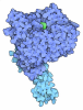
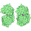
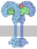
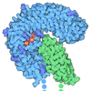








- Assembly
| Deposited unit | 
|
|---|---|
| 1 |
|
-Components
-Protein , 5 types, 5 molecules ABCDR
| #1: Protein | Mass: 84463.344 Da / Num. of mol.: 1 Source method: isolated from a genetically manipulated source Source: (gene. exp.) Homo sapiens (human) / Gene: ZYG11B, KIAA1730 / Production host:   Escherichia coli (E. coli) / References: UniProt: Q9C0D3 Escherichia coli (E. coli) / References: UniProt: Q9C0D3 |
|---|---|
| #2: Protein | Mass: 13147.781 Da / Num. of mol.: 1 Source method: isolated from a genetically manipulated source Source: (gene. exp.) Homo sapiens (human) / Gene: ELOB, TCEB2 / Production host: Escherichia coli (E. coli) / References: UniProt: Q15370 |
| #3: Protein | Mass: 10843.420 Da / Num. of mol.: 1 Source method: isolated from a genetically manipulated source Source: (gene. exp.) Homo sapiens (human) / Gene: ELOC, TCEB1 / Production host: Escherichia coli (E. coli) / References: UniProt: Q15369 |
| #4: Protein | Mass: 86967.734 Da / Num. of mol.: 1 Source method: isolated from a genetically manipulated source Source: (gene. exp.) Homo sapiens (human) / Gene: CUL2 / Production host:   Spodoptera frugiperda (fall armyworm) / References: UniProt: Q13617 Spodoptera frugiperda (fall armyworm) / References: UniProt: Q13617 |
| #5: Protein | Mass: 12289.977 Da / Num. of mol.: 1 Source method: isolated from a genetically manipulated source Source: (gene. exp.) Homo sapiens (human) / Gene: RBX1, RNF75, ROC1 / Production host: Spodoptera frugiperda (fall armyworm)References: UniProt: P62877, RING-type E3 ubiquitin transferase, cullin-RING-type E3 NEDD8 transferase |
-Non-polymers , 1 types, 3 molecules 
| #6: Chemical | 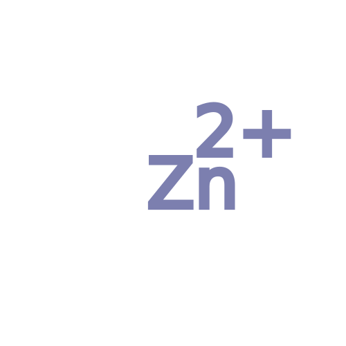 Mass: 65.409 Da / Num. of mol.: 3 / Source method: obtained synthetically / Formula: Zn / Feature type: SUBJECT OF INVESTIGATION Mass: 65.409 Da / Num. of mol.: 3 / Source method: obtained synthetically / Formula: Zn / Feature type: SUBJECT OF INVESTIGATION |
|---|
-Details
| Has ligand of interest | Y |
|---|---|
| Has protein modification | N |
-Experimental details
-Experiment
| Experiment | Method: ELECTRON MICROSCOPY |
|---|---|
| EM experiment | Aggregation state: PARTICLE / 3D reconstruction method: single particle reconstruction |
- Sample preparation
| Component |
| ||||||||||||||||||||||||||||||
|---|---|---|---|---|---|---|---|---|---|---|---|---|---|---|---|---|---|---|---|---|---|---|---|---|---|---|---|---|---|---|---|
| Molecular weight | Value: 230 kDa/nm / Experimental value: NO | ||||||||||||||||||||||||||||||
| Source (natural) |
| ||||||||||||||||||||||||||||||
| Source (recombinant) |
| ||||||||||||||||||||||||||||||
| Buffer solution | pH: 7.2 | ||||||||||||||||||||||||||||||
| Specimen | Embedding applied: NO / Shadowing applied: NO / Staining applied: NO / Vitrification applied: YES | ||||||||||||||||||||||||||||||
| Specimen support | Grid material: GOLD / Grid mesh size: 300 divisions/in. / Grid type: Quantifoil R1.2/1.3 | ||||||||||||||||||||||||||||||
| Vitrification | Cryogen name: ETHANE |
- Electron microscopy imaging
| Experimental equipment |  Model: Titan Krios / Image courtesy: FEI Company |
|---|---|
| Microscopy | Model: FEI TITAN KRIOS |
| Electron gun | Electron source:  FIELD EMISSION GUN / Accelerating voltage: 300 kV / Illumination mode: FLOOD BEAM FIELD EMISSION GUN / Accelerating voltage: 300 kV / Illumination mode: FLOOD BEAM |
| Electron lens | Mode: 4D-STEM / Nominal defocus max: 5000 nm / Nominal defocus min: 1000 nm |
| Specimen holder | Cryogen: NITROGEN / Specimen holder model: FEI TITAN KRIOS AUTOGRID HOLDER |
| Image recording | Electron dose: 60 e/Å2 / Film or detector model: GATAN K3 (6k x 4k) |
- Processing
| EM software |
| ||||||||||||||||||||||||
|---|---|---|---|---|---|---|---|---|---|---|---|---|---|---|---|---|---|---|---|---|---|---|---|---|---|
| CTF correction | Type: NONE | ||||||||||||||||||||||||
| 3D reconstruction | Resolution: 3.78 Å / Resolution method: FSC 0.143 CUT-OFF / Num. of particles: 177561 / Algorithm: ALGEBRAIC (ARTS) / Symmetry type: POINT | ||||||||||||||||||||||||
| Refinement | Cross valid method: NONE Stereochemistry target values: GeoStd + Monomer Library + CDL v1.2 | ||||||||||||||||||||||||
| Displacement parameters | Biso mean: 127.87 Å2 | ||||||||||||||||||||||||
| Refine LS restraints |
|
