Movie
Movie Controller

[English] 日本語
 Yorodumi
- PDB-8yt4: Structure of Aquifex aeolicus Lumazine Synthase by Cryo-Electron ... -
+ Open data

- Basic information
| Entry | Database: PDB / ID: 8yt4 | ||||||
|---|---|---|---|---|---|---|---|
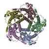
| Title | Structure of Aquifex aeolicus Lumazine Synthase by Cryo-Electron Microscopy to 1.42 Angstrom Resolution | ||||||
 Components Components | 6,7-dimethyl-8-ribityllumazine synthase | ||||||
 Keywords Keywords | BIOSYNTHETIC PROTEIN / Enzyme involved in riboflavin biosynthesis | ||||||
| Function / homology |  Function and homology information Function and homology information6,7-dimethyl-8-ribityllumazine synthase / 6,7-dimethyl-8-ribityllumazine synthase activity / riboflavin synthase complex / riboflavin biosynthetic process / cytoplasm / cytosol Similarity search - Function | ||||||
| Biological species |   Aquifex aeolicus (bacteria) Aquifex aeolicus (bacteria) | ||||||
| Method | ELECTRON MICROSCOPY / single particle reconstruction / cryo EM / Resolution: 1.42 Å | ||||||
 Authors Authors | Savva, C.G. / Sobhy, M.A. / De Biasio, A. / Hamdan, S.M. | ||||||
| Funding support | 1items
| ||||||
 Citation Citation | Journal: IUCrJ / Year: 2024 Title: Structure of Aquifex aeolicus lumazine synthase by cryo-electron microscopy to 1.42 Å resolution. Authors: Christos G Savva / Mohamed A Sobhy / Alfredo De Biasio / Samir M Hamdan /  Abstract: Single-particle cryo-electron microscopy (cryo-EM) has become an essential structural determination technique with recent hardware developments making it possible to reach atomic resolution, at which ...Single-particle cryo-electron microscopy (cryo-EM) has become an essential structural determination technique with recent hardware developments making it possible to reach atomic resolution, at which individual atoms, including hydrogen atoms, can be resolved. In this study, we used the enzyme involved in the penultimate step of riboflavin biosynthesis as a test specimen to benchmark a recently installed microscope and determine if other protein complexes could reach a resolution of 1.5 Å or better, which so far has only been achieved for the iron carrier ferritin. Using state-of-the-art microscope and detector hardware as well as the latest software techniques to overcome microscope and sample limitations, a 1.42 Å map of Aquifex aeolicus lumazine synthase (AaLS) was obtained from a 48 h microscope session. In addition to water molecules and ligands involved in the function of AaLS, we can observe positive density for ∼50% of the hydrogen atoms. A small improvement in the resolution was achieved by Ewald sphere correction which was expected to limit the resolution to ∼1.5 Å for a molecule of this diameter. Our study confirms that other protein complexes can be solved to near-atomic resolution. Future improvements in specimen preparation and protein complex stabilization may allow more flexible macromolecules to reach this level of resolution and should become a priority of study in the field. | ||||||
| History |
|
- Structure visualization
| Structure viewer | Molecule: MolmilJmol/JSmol |
|---|
- Downloads & links
-Download
| PDBx/mmCIF format | 8yt4.cif.gz | 59.4 KB | Display | PDBx/mmCIF format |
|---|---|---|---|---|
| PDB format | pdb8yt4.ent.gz | 39.9 KB | Display | PDB format |
| PDBx/mmJSON format | 8yt4.json.gz | Tree view | PDBx/mmJSON format | |
| Others |  Other downloads Other downloads |
-Validation report
| Arichive directory | https://data.pdbj.org/pub/pdb/validation_reports/yt/8yt4ftp://data.pdbj.org/pub/pdb/validation_reports/yt/8yt4 | HTTPS FTP |
|---|
-Related structure data
| Related structure data |  39478MC M: map data used to model this data C: citing same article ( |
|---|---|
| Similar structure data |
-Links
 PDBj
PDBj- Assembly
| Deposited unit | 
|
|---|---|
| 1 | x 60
|
| 2 |
|
| 3 | x 5
|
| 4 | x 6
|
| 5 | 
|
| Symmetry | Point symmetry: (Schoenflies symbol: I (icosahedral)) |
-Components
| #1: Protein | Mass: 17769.350 Da / Num. of mol.: 1 Source method: isolated from a genetically manipulated source Source: (gene. exp.) Aquifex aeolicus (bacteria) / Gene: ribH, aq_132 / Production host: References: UniProt: O66529, 6,7-dimethyl-8-ribityllumazine synthase |
|---|---|
| #2: Chemical | ChemComp-PO4 /   Mass: 94.971 Da / Num. of mol.: 1 / Source method: obtained synthetically / Formula: PO4 / Feature type: SUBJECT OF INVESTIGATION Mass: 94.971 Da / Num. of mol.: 1 / Source method: obtained synthetically / Formula: PO4 / Feature type: SUBJECT OF INVESTIGATION |
| #3: Water | ChemComp-HOH /  Mass: 18.015 Da / Num. of mol.: 55 / Source method: isolated from a natural source / Formula: H2O Mass: 18.015 Da / Num. of mol.: 55 / Source method: isolated from a natural source / Formula: H2O |
| Has ligand of interest | Y |
-Experimental details
-Experiment
| Experiment | Method: ELECTRON MICROSCOPY |
|---|---|
| EM experiment | Aggregation state: PARTICLE / 3D reconstruction method: single particle reconstruction |
- Sample preparation
| Component | Name: Lumazine synthase / Type: COMPLEX / Entity ID: #1 / Source: RECOMBINANT |
|---|---|
| Molecular weight | Value: 1.065 MDa / Experimental value: NO |
| Source (natural) | Organism: Aquifex aeolicus (bacteria) |
| Source (recombinant) | Organism: |
| Buffer solution | pH: 7.5 / Details: 20mM Tris pH7.5, 150mM NaCl, 1 mM DTT |
| Specimen | Conc.: 2.75 mg/ml / Embedding applied: NO / Shadowing applied: NO / Staining applied: NO / Vitrification applied: YES |
| Specimen support | Details: 3O mA / Grid material: GOLD / Grid mesh size: 300 divisions/in. / Grid type: UltrAuFoil R1.2/1.3 |
| Vitrification | Instrument: FEI VITROBOT MARK IV / Cryogen name: ETHANE / Humidity: 100 % / Chamber temperature: 277 K |
- Electron microscopy imaging
| Experimental equipment |  Model: Titan Krios / Image courtesy: FEI Company |
|---|---|
| Microscopy | Model: FEI TITAN KRIOS |
| Electron gun | Electron source:  FIELD EMISSION GUN / Accelerating voltage: 300 kV / Illumination mode: FLOOD BEAM FIELD EMISSION GUN / Accelerating voltage: 300 kV / Illumination mode: FLOOD BEAM |
| Electron lens | Mode: BRIGHT FIELD / Nominal magnification: 270000 X / Nominal defocus max: 1200 nm / Nominal defocus min: 400 nm / Cs: 2.7 mm / C2 aperture diameter: 50 µm / Alignment procedure: COMA FREE |
| Specimen holder | Cryogen: NITROGEN / Specimen holder model: FEI TITAN KRIOS AUTOGRID HOLDER / Temperature (min): 85 K |
| Image recording | Average exposure time: 3 sec. / Electron dose: 46 e/Å2 / Film or detector model: FEI FALCON IV (4k x 4k) / Num. of grids imaged: 1 / Num. of real images: 12657 |
| EM imaging optics | Energyfilter name: TFS Selectris X / Energyfilter slit width: 10 eV |
| Image scans | Width: 4096 / Height: 4096 |
- Processing
| EM software |
| ||||||||||||||||||||||||||||||||||||||||||||||||||||||||||||||||||||||||||||||||||||||||||||||||||||||||||
|---|---|---|---|---|---|---|---|---|---|---|---|---|---|---|---|---|---|---|---|---|---|---|---|---|---|---|---|---|---|---|---|---|---|---|---|---|---|---|---|---|---|---|---|---|---|---|---|---|---|---|---|---|---|---|---|---|---|---|---|---|---|---|---|---|---|---|---|---|---|---|---|---|---|---|---|---|---|---|---|---|---|---|---|---|---|---|---|---|---|---|---|---|---|---|---|---|---|---|---|---|---|---|---|---|---|---|---|
| CTF correction | Type: PHASE FLIPPING AND AMPLITUDE CORRECTION | ||||||||||||||||||||||||||||||||||||||||||||||||||||||||||||||||||||||||||||||||||||||||||||||||||||||||||
| Particle selection | Num. of particles selected: 698240 | ||||||||||||||||||||||||||||||||||||||||||||||||||||||||||||||||||||||||||||||||||||||||||||||||||||||||||
| Symmetry | Point symmetry: I (icosahedral) | ||||||||||||||||||||||||||||||||||||||||||||||||||||||||||||||||||||||||||||||||||||||||||||||||||||||||||
| 3D reconstruction | Resolution: 1.42 Å / Resolution method: FSC 0.143 CUT-OFF / Num. of particles: 470878 / Algorithm: BACK PROJECTION / Symmetry type: POINT | ||||||||||||||||||||||||||||||||||||||||||||||||||||||||||||||||||||||||||||||||||||||||||||||||||||||||||
| Atomic model building | Protocol: RIGID BODY FIT | ||||||||||||||||||||||||||||||||||||||||||||||||||||||||||||||||||||||||||||||||||||||||||||||||||||||||||
| Atomic model building | PDB-ID: 1HQK Pdb chain-ID: A / Accession code: 1HQK / Source name: PDB / Type: experimental model | ||||||||||||||||||||||||||||||||||||||||||||||||||||||||||||||||||||||||||||||||||||||||||||||||||||||||||
| Refinement | Resolution: 1.42→1.42 Å / Cor.coef. Fo:Fc: 0.669 / SU B: 0.611 / SU ML: 0.023 / ESU R: 0.006 Stereochemistry target values: MAXIMUM LIKELIHOOD WITH PHASES Details: HYDROGENS HAVE BEEN USED IF PRESENT IN THE INPUT
| ||||||||||||||||||||||||||||||||||||||||||||||||||||||||||||||||||||||||||||||||||||||||||||||||||||||||||
| Solvent computation | Solvent model: PARAMETERS FOR MASK CACLULATION | ||||||||||||||||||||||||||||||||||||||||||||||||||||||||||||||||||||||||||||||||||||||||||||||||||||||||||
| Displacement parameters | Biso mean: 20.034 Å2 | ||||||||||||||||||||||||||||||||||||||||||||||||||||||||||||||||||||||||||||||||||||||||||||||||||||||||||
| Refinement step | Cycle: 1 / Total: 1225 | ||||||||||||||||||||||||||||||||||||||||||||||||||||||||||||||||||||||||||||||||||||||||||||||||||||||||||
| Refine LS restraints |
|