 Movie
Movie Controller

[English] 日本語
 Yorodumi
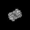
- PDB-8yf5: Cryo EM structure of Komagataella phaffii Rat1-Rai1-Rtt103 complex -
+ Open data

- Basic information
| Entry | Database: PDB / ID: 8yf5 | ||||||
|---|---|---|---|---|---|---|---|
| Title | Cryo EM structure of Komagataella phaffii Rat1-Rai1-Rtt103 complex | ||||||
 Components Components |
| ||||||
 Keywords Keywords | TRANSCRIPTION / transcription termination / RNA polymerase II | ||||||
| Function / homology |  Function and homology information Function and homology informationmRNA 5'-diphosphatase activity / NAD-cap decapping / 5'-3' RNA exonuclease activity / nuclease activity / mRNA 3'-end processing / exonuclease activity / Hydrolases; Acting on acid anhydrides; In phosphorus-containing anhydrides / nuclear-transcribed mRNA catabolic process / RNA polymerase II C-terminal domain binding / DNA-templated transcription termination ...mRNA 5'-diphosphatase activity / NAD-cap decapping / 5'-3' RNA exonuclease activity / nuclease activity / mRNA 3'-end processing / exonuclease activity / Hydrolases; Acting on acid anhydrides; In phosphorus-containing anhydrides / nuclear-transcribed mRNA catabolic process / RNA polymerase II C-terminal domain binding / DNA-templated transcription termination / mRNA processing / rRNA processing / Hydrolases; Acting on ester bonds; Exoribonucleases producing 5'-phosphomonoesters / nucleic acid binding / nucleotide binding / RNA binding / nucleus / metal ion binding / cytosol Similarity search - Function | ||||||
| Biological species |  Komagataella phaffii (fungus) Komagataella phaffii (fungus) | ||||||
| Method | ELECTRON MICROSCOPY / single particle reconstruction / cryo EM / Resolution: 3.78 Å | ||||||
 Authors Authors | Yanagisawa, T. / Murayama, Y. / Ehara, H. / Sekine, S.I. | ||||||
| Funding support |  Japan, 1items Japan, 1items
| ||||||
 Citation Citation | Journal: Nat Commun / Year: 2024 Title: Structural basis of eukaryotic transcription termination by the Rat1 exonuclease complex. Authors: Tatsuo Yanagisawa / Yuko Murayama / Haruhiko Ehara / Mie Goto / Mari Aoki / Shun-Ichi Sekine / Abstract: The 5´-3´ exoribonuclease Rat1/Xrn2 is responsible for the termination of eukaryotic mRNA transcription by RNAPII. Rat1 forms a complex with its partner proteins, Rai1 and Rtt103, and acts as a ...The 5´-3´ exoribonuclease Rat1/Xrn2 is responsible for the termination of eukaryotic mRNA transcription by RNAPII. Rat1 forms a complex with its partner proteins, Rai1 and Rtt103, and acts as a "torpedo" to bind transcribing RNAPII and dissociate DNA/RNA from it. Here we report the cryo-electron microscopy structures of the Rat1-Rai1-Rtt103 complex and three Rat1-Rai1-associated RNAPII complexes (type-1, type-1b, and type-2) from the yeast, Komagataella phaffii. The Rat1-Rai1-Rtt103 structure revealed that Rat1 and Rai1 form a heterotetramer with a single Rtt103 bound between two Rai1 molecules. In the type-1 complex, Rat1-Rai1 forms a heterodimer and binds to the RNA exit site of RNAPII to extract RNA into the Rat1 exonuclease active site. This interaction changes the RNA path in favor of termination (the "pre-termination" state). The type-1b and type-2 complexes have no bound DNA/RNA, likely representing the "post-termination" states. These structures illustrate the termination mechanism of eukaryotic mRNA transcription. | ||||||
| History |
|
- Structure visualization
| Structure viewer | Molecule: MolmilJmol/JSmol |
|---|
- Downloads & links
-Download
| PDBx/mmCIF format | 8yf5.cif.gz | 426.7 KB | Display | PDBx/mmCIF format |
|---|---|---|---|---|
| PDB format | pdb8yf5.ent.gz | 336.6 KB | Display | PDB format |
| PDBx/mmJSON format | 8yf5.json.gz | Tree view | PDBx/mmJSON format | |
| Others |  Other downloads Other downloads |
-Validation report
| Summary document | 8yf5_validation.pdf.gz | 1.2 MB | Display | wwPDB validaton report |
|---|---|---|---|---|
| Full document | 8yf5_full_validation.pdf.gz | 1.3 MB | Display | |
| Data in XML | 8yf5_validation.xml.gz | 66.7 KB | Display | |
| Data in CIF | 8yf5_validation.cif.gz | 102.3 KB | Display | |
| Arichive directory | https://data.pdbj.org/pub/pdb/validation_reports/yf/8yf5ftp://data.pdbj.org/pub/pdb/validation_reports/yf/8yf5 | HTTPS FTP |
-Related structure data
| Related structure data |  39211MC 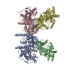 8yfeC 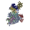 8yfqC 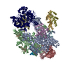 8yfrC M: map data used to model this data C: citing same article ( |
|---|---|
| Similar structure data |
-Links
 PDBj
PDBj
- Assembly
| Deposited unit | 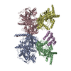
|
|---|---|
| 1 |
|
-Components
| #1: Protein | Mass: 115314.461 Da / Num. of mol.: 2 / Mutation: D233A, D235A Source method: isolated from a genetically manipulated source Source: (gene. exp.) Komagataella phaffii (fungus) / Gene: RAT1, PP7435_Chr3-0338 / Production host:  References: UniProt: F2QV79, Hydrolases; Acting on ester bonds; Exoribonucleases producing 5'-phosphomonoesters #2: Protein | Mass: 44595.996 Da / Num. of mol.: 2 / Mutation: E213A, D215A Source method: isolated from a genetically manipulated source Source: (gene. exp.) Komagataella phaffii (fungus) / Gene: RAI1, PP7435_Chr1-1057 / Production host: References: UniProt: F2QLF5, Hydrolases; Acting on acid anhydrides; In phosphorus-containing anhydrides #3: Protein | | Mass: 42272.930 Da / Num. of mol.: 1 Source method: isolated from a genetically manipulated source Source: (gene. exp.) Komagataella phaffii (fungus) / Gene: RTT103, PP7435_Chr1-0928 / Production host: |
|---|
-Experimental details
-Experiment
| Experiment | Method: ELECTRON MICROSCOPY |
|---|---|
| EM experiment | Aggregation state: PARTICLE / 3D reconstruction method: single particle reconstruction |
- Sample preparation
| Component | Name: Komagataella phaffii Rat1-Rai1-Rtt103 complex / Type: COMPLEX / Entity ID: all / Source: RECOMBINANT | ||||||||||||||||||||||||||||||
|---|---|---|---|---|---|---|---|---|---|---|---|---|---|---|---|---|---|---|---|---|---|---|---|---|---|---|---|---|---|---|---|
| Molecular weight | Experimental value: NO | ||||||||||||||||||||||||||||||
| Source (natural) | Organism: Komagataella phaffii (fungus) | ||||||||||||||||||||||||||||||
| Source (recombinant) | Organism: | ||||||||||||||||||||||||||||||
| Buffer solution | pH: 7.5 | ||||||||||||||||||||||||||||||
| Buffer component |
| ||||||||||||||||||||||||||||||
| Specimen | Embedding applied: NO / Shadowing applied: NO / Staining applied: NO / Vitrification applied: YES | ||||||||||||||||||||||||||||||
| Specimen support | Grid material: COPPER / Grid mesh size: 300 divisions/in. / Grid type: Quantifoil R1.2/1.3 | ||||||||||||||||||||||||||||||
| Vitrification | Instrument: LEICA EM GP / Cryogen name: ETHANE / Humidity: 75 % / Chamber temperature: 283 K / Details: EMGP2 |
- Electron microscopy imaging
| Experimental equipment |  Model: Titan Krios / Image courtesy: FEI Company |
|---|---|
| Microscopy | Model: TFS KRIOS |
| Electron gun | Electron source:  FIELD EMISSION GUN / Accelerating voltage: 300 kV / Illumination mode: FLOOD BEAM FIELD EMISSION GUN / Accelerating voltage: 300 kV / Illumination mode: FLOOD BEAM |
| Electron lens | Mode: BRIGHT FIELD / Nominal defocus max: 2400 nm / Nominal defocus min: 1600 nm |
| Image recording | Electron dose: 61.9 e/Å2 / Film or detector model: GATAN K3 (6k x 4k) |
- Processing
| CTF correction | Type: NONE | ||||||||||||||||||||||||
|---|---|---|---|---|---|---|---|---|---|---|---|---|---|---|---|---|---|---|---|---|---|---|---|---|---|
| 3D reconstruction | Resolution: 3.78 Å / Resolution method: FSC 0.143 CUT-OFF / Num. of particles: 77924 / Symmetry type: POINT | ||||||||||||||||||||||||
| Refine LS restraints |
|
