 Movie
Movie Controller


+ Open data

- Basic information
| Entry | Database: PDB / ID: 8xj4 | |||||||||||||||||||||||||||||||||||||||||||||||||||||||||||||||
|---|---|---|---|---|---|---|---|---|---|---|---|---|---|---|---|---|---|---|---|---|---|---|---|---|---|---|---|---|---|---|---|---|---|---|---|---|---|---|---|---|---|---|---|---|---|---|---|---|---|---|---|---|---|---|---|---|---|---|---|---|---|---|---|---|
| Title | Structure of prostatic acid phosphatase in human semen | |||||||||||||||||||||||||||||||||||||||||||||||||||||||||||||||
 Components Components | Prostatic acid phosphatase | |||||||||||||||||||||||||||||||||||||||||||||||||||||||||||||||
 Keywords Keywords | CELL CYCLE / Acid phosphatase | |||||||||||||||||||||||||||||||||||||||||||||||||||||||||||||||
| Function / homology |  Function and homology information Function and homology informationthiamine phosphate phosphatase activity / positive regulation of adenosine receptor signaling pathway / thiamine metabolic process / Golgi cisterna / adenosine metabolic process / acid phosphatase / acid phosphatase activity / regulation of sensory perception of pain / lysophosphatidic acid phosphatase activity / 5'-nucleotidase ...thiamine phosphate phosphatase activity / positive regulation of adenosine receptor signaling pathway / thiamine metabolic process / Golgi cisterna / adenosine metabolic process / acid phosphatase / acid phosphatase activity / regulation of sensory perception of pain / lysophosphatidic acid phosphatase activity / 5'-nucleotidase / choline binding / 5'-nucleotidase activity / nucleotide metabolic process / vesicle membrane / azurophil granule membrane / purine nucleobase metabolic process / phosphatase activity / multivesicular body / protein-tyrosine-phosphatase / protein tyrosine phosphatase activity / filopodium / lipid metabolic process / apical part of cell / molecular adaptor activity / lysosomal membrane / Neutrophil degranulation / protein homodimerization activity / : / extracellular exosome / identical protein binding / nucleus / plasma membrane / cytosol Similarity search - Function | |||||||||||||||||||||||||||||||||||||||||||||||||||||||||||||||
| Biological species |  Homo sapiens (human) Homo sapiens (human) | |||||||||||||||||||||||||||||||||||||||||||||||||||||||||||||||
| Method | ELECTRON MICROSCOPY / single particle reconstruction / cryo EM / Resolution: 3.19 Å | |||||||||||||||||||||||||||||||||||||||||||||||||||||||||||||||
 Authors Authors | Liu, X.Z. / Li, J.L. / Deng, D. / Wang, X. | |||||||||||||||||||||||||||||||||||||||||||||||||||||||||||||||
| Funding support |  China, 1items China, 1items
| |||||||||||||||||||||||||||||||||||||||||||||||||||||||||||||||
 Citation Citation | Journal: Biochem Biophys Res Commun / Year: 2024 Title: Purification, identification and Cryo-EM structure of prostatic acid phosphatase in human semen. Authors: Xuanzhong Liu / Lin Yu / Zhili Xia / Jialu Li / Wenbo Meng / Ling Min / Fuping Li / Xiang Wang / Abstract: Prostatic acid phosphatase (PAP) is a glycoprotein that plays a crucial role in the hydrolysis of phosphate ester present in prostatic exudates. It is a well-established indicator for prostate cancer ...Prostatic acid phosphatase (PAP) is a glycoprotein that plays a crucial role in the hydrolysis of phosphate ester present in prostatic exudates. It is a well-established indicator for prostate cancer due to its elevated serum levels in disease progression. Despite its abundance in semen, PAP's influence on male fertility has not been extensively studied. In our study, we report a significantly optimized method for purifying human endogenous PAP, achieving remarkably high efficiency and active protein recovery rate. This achievement allowed us to better analyze and understand the PAP protein. We determined the cryo-electron microscopic (Cryo-EM) structure of prostatic acid phosphatase in its physiological state for the first time. Our structural and gel filtration analysis confirmed the formation of a tight homodimer structure of human PAP. This functional homodimer displayed an elongated conformation in the cryo-EM structure compared to the previously reported crystal structure. Additionally, there was a notable 5-degree rotation in the angle between the α domain and α/β domain of each monomer. Through structural analysis, we revealed three potential glycosylation sites: Asn94, Asn220, and Asn333. These sites contained varying numbers and forms of glycosyl units, suggesting sugar moieties influence PAP function. Furthermore, we found that the active sites of PAP, His44 and Asp290, are located between the two protein domains. Overall, our study not only provide an optimized approach for PAP purification, but also offer crucial insights into its structural characteristics. These findings lay the groundwork for further investigations into the physiological function and potential therapeutic applications of this important protein. | |||||||||||||||||||||||||||||||||||||||||||||||||||||||||||||||
| History |
|
- Structure visualization
| Structure viewer | Molecule: MolmilJmol/JSmol |
|---|
- Downloads & links
-Download
| PDBx/mmCIF format | 8xj4.cif.gz | 139.2 KB | Display | PDBx/mmCIF format |
|---|---|---|---|---|
| PDB format | pdb8xj4.ent.gz | 107.9 KB | Display | PDB format |
| PDBx/mmJSON format | 8xj4.json.gz | Tree view | PDBx/mmJSON format | |
| Others |  Other downloads Other downloads |
-Validation report
| Arichive directory | https://data.pdbj.org/pub/pdb/validation_reports/xj/8xj4ftp://data.pdbj.org/pub/pdb/validation_reports/xj/8xj4 | HTTPS FTP |
|---|
-Related structure data
| Related structure data | 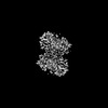 38393MC M: map data used to model this data C: citing same article ( |
|---|---|
| Similar structure data |
-Links
 PDBj
PDBj


- Assembly
| Deposited unit | 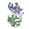
|
|---|---|
| 1 |
|
-Components
| #1: Protein | Mass: 40262.949 Da / Num. of mol.: 2 / Source method: isolated from a natural source / Source: (natural) Homo sapiens (human) / Tissue: Human seminal plasmaReferences: UniProt: P15309, acid phosphatase, 5'-nucleotidase, protein-tyrosine-phosphatase #2: Polysaccharide | alpha-D-mannopyranose-(1-3)-2-acetamido-2-deoxy-beta-D-glucopyranose-(1-4)-2-acetamido-2-deoxy-beta- ...alpha-D-mannopyranose-(1-3)-2-acetamido-2-deoxy-beta-D-glucopyranose-(1-4)-2-acetamido-2-deoxy-beta-D-glucopyranose | #3: Polysaccharide | alpha-D-mannopyranose-(1-2)-alpha-D-mannopyranose-(1-3)-2-acetamido-2-deoxy-beta-D-glucopyranose-(1- ...alpha-D-mannopyranose-(1-2)-alpha-D-mannopyranose-(1-3)-2-acetamido-2-deoxy-beta-D-glucopyranose-(1-4)-2-acetamido-2-deoxy-beta-D-glucopyranose | Type: oligosaccharide / Mass: 748.682 Da / Num. of mol.: 1 / Source method: obtained synthetically #4: Sugar | ChemComp-NAG /   Type: D-saccharide, beta linking / Mass: 221.208 Da / Num. of mol.: 4 / Source method: obtained synthetically / Formula: C8H15NO6 / Feature type: SUBJECT OF INVESTIGATION Type: D-saccharide, beta linking / Mass: 221.208 Da / Num. of mol.: 4 / Source method: obtained synthetically / Formula: C8H15NO6 / Feature type: SUBJECT OF INVESTIGATION#5: Sugar | ChemComp-MAN / |   Type: D-saccharide, alpha linking / Mass: 180.156 Da / Num. of mol.: 1 / Source method: obtained synthetically / Formula: C6H12O6 / Feature type: SUBJECT OF INVESTIGATION Type: D-saccharide, alpha linking / Mass: 180.156 Da / Num. of mol.: 1 / Source method: obtained synthetically / Formula: C6H12O6 / Feature type: SUBJECT OF INVESTIGATIONHas ligand of interest | Y | Has protein modification | Y | |
|---|
-Experimental details
-Experiment
| Experiment | Method: ELECTRON MICROSCOPY |
|---|---|
| EM experiment | Aggregation state: PARTICLE / 3D reconstruction method: single particle reconstruction |
- Sample preparation
| Component | Name: structure of prostatic acid phosphatase in human semen Type: ORGANELLE OR CELLULAR COMPONENT / Entity ID: #1 / Source: NATURAL |
|---|---|
| Source (natural) | Organism: Homo sapiens (human) |
| Source (recombinant) | Organism:   Spodoptera frugiperda (fall armyworm) Spodoptera frugiperda (fall armyworm) |
| Buffer solution | pH: 8 |
| Specimen | Conc.: 8.5 mg/ml / Embedding applied: NO / Shadowing applied: NO / Staining applied: NO / Vitrification applied: YES |
| Vitrification | Cryogen name: ETHANE |
- Electron microscopy imaging
| Experimental equipment |  Model: Titan Krios / Image courtesy: FEI Company |
|---|---|
| Microscopy | Model: FEI TITAN KRIOS |
| Electron gun | Electron source:  FIELD EMISSION GUN / Accelerating voltage: 300 kV / Illumination mode: FLOOD BEAM FIELD EMISSION GUN / Accelerating voltage: 300 kV / Illumination mode: FLOOD BEAM |
| Electron lens | Mode: BRIGHT FIELD / Nominal defocus max: 1800 nm / Nominal defocus min: 1200 nm |
| Image recording | Electron dose: 50 e/Å2 / Film or detector model: GATAN K2 SUMMIT (4k x 4k) |
- Processing
| EM software | Name: PHENIX / Category: model refinement | ||||||||||||||||||||||||
|---|---|---|---|---|---|---|---|---|---|---|---|---|---|---|---|---|---|---|---|---|---|---|---|---|---|
| CTF correction | Type: NONE | ||||||||||||||||||||||||
| 3D reconstruction | Resolution: 3.19 Å / Resolution method: FSC 0.143 CUT-OFF / Num. of particles: 75346 / Symmetry type: POINT | ||||||||||||||||||||||||
| Refine LS restraints |
|
