 Movie
Movie Controller


+ Open data

- Basic information
| Entry | Database: PDB / ID: 8xh7 | ||||||
|---|---|---|---|---|---|---|---|
| Title | Structure of EBV LMP1 oligomer | ||||||
 Components Components | Latent membrane protein 1 | ||||||
 Keywords Keywords | VIRAL PROTEIN / EBV latent membrane protein 1 | ||||||
| Function / homology |  Function and homology information Function and homology informationsymbiont-mediated activation of host NF-kappaB cascade / symbiont-mediated transformation of host cell / symbiont-mediated suppression of host TRAF-mediated signal transduction / symbiont-mediated suppression of host JAK-STAT cascade via inhibition of host TYK2 activity / symbiont-mediated suppression of host type I interferon-mediated signaling pathway / host cell plasma membrane / membrane Similarity search - Function | ||||||
| Biological species |  human gammaherpesvirus 4 (Epstein-Barr virus) human gammaherpesvirus 4 (Epstein-Barr virus) | ||||||
| Method | ELECTRON MICROSCOPY / single particle reconstruction / cryo EM / Resolution: 3.52 Å | ||||||
 Authors Authors | Gao, P. / Huang, J.F. | ||||||
| Funding support | 1items
| ||||||
 Citation Citation | Journal: Cell / Year: 2024 Title: Assembly and activation of EBV latent membrane protein 1. Authors: Jiafeng Huang / Xiaolin Zhang / Xiaohua Nie / Xuyuan Zhang / Yong Wang / Linlong Huang / Xiaohan Geng / Dong Li / Liguo Zhang / Guangxia Gao / Pu Gao /  Abstract: Latent membrane protein 1 (LMP1) is the primary oncoprotein of Epstein-Barr virus (EBV) and plays versatile roles in the EBV life cycle and pathogenesis. Despite decades of extensive research, the ...Latent membrane protein 1 (LMP1) is the primary oncoprotein of Epstein-Barr virus (EBV) and plays versatile roles in the EBV life cycle and pathogenesis. Despite decades of extensive research, the molecular basis for LMP1 folding, assembly, and activation remains unclear. Here, we report cryo-electron microscopy structures of LMP1 in two unexpected assemblies: a symmetric homodimer and a higher-order filamentous oligomer. LMP1 adopts a non-canonical and unpredicted fold that supports the formation of a stable homodimer through tight and antiparallel intermolecular packing. LMP1 dimers further assemble side-by-side into higher-order filamentous oligomers, thereby allowing the accumulation and specific organization of the flexible cytoplasmic tails for efficient recruitment of downstream factors. Super-resolution microscopy and cellular functional assays demonstrate that mutations at both dimeric and oligomeric interfaces disrupt LMP1 higher-order assembly and block multiple LMP1-mediated signaling pathways. Our research provides a framework for understanding the mechanism of LMP1 and for developing potential therapies targeting EBV-associated diseases. | ||||||
| History |
|
- Structure visualization
| Structure viewer | Molecule: MolmilJmol/JSmol |
|---|
- Downloads & links
-Download
| PDBx/mmCIF format | 8xh7.cif.gz | 172.6 KB | Display | PDBx/mmCIF format |
|---|---|---|---|---|
| PDB format | pdb8xh7.ent.gz | 140.9 KB | Display | PDB format |
| PDBx/mmJSON format | 8xh7.json.gz | Tree view | PDBx/mmJSON format | |
| Others |  Other downloads Other downloads |
-Validation report
| Arichive directory | https://data.pdbj.org/pub/pdb/validation_reports/xh/8xh7ftp://data.pdbj.org/pub/pdb/validation_reports/xh/8xh7 | HTTPS FTP |
|---|
-Related structure data
| Related structure data |  38343MC  8xh6C M: map data used to model this data C: citing same article ( |
|---|---|
| Similar structure data |
-Links
 PDBj
PDBj- Assembly
| Deposited unit | 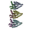
|
|---|---|
| 1 |
|
-Components
| #1: Protein | Mass: 18729.418 Da / Num. of mol.: 6 Source method: isolated from a genetically manipulated source Source: (gene. exp.) human gammaherpesvirus 4 (Epstein-Barr virus)Gene: LMP1, LMP-1 / Production host:  |
|---|
-Experimental details
-Experiment
| Experiment | Method: ELECTRON MICROSCOPY |
|---|---|
| EM experiment | Aggregation state: PARTICLE / 3D reconstruction method: single particle reconstruction |
- Sample preparation
| Component | Name: LMP1 oligomer / Type: COMPLEX / Entity ID: all / Source: RECOMBINANT |
|---|---|
| Source (natural) | Organism: Human gammaherpesvirus 4 (Epstein-Barr virus) |
| Source (recombinant) | Organism: |
| Buffer solution | pH: 7.5 |
| Specimen | Embedding applied: NO / Shadowing applied: NO / Staining applied: NO / Vitrification applied: YES |
| Vitrification | Cryogen name: ETHANE |
- Electron microscopy imaging
| Experimental equipment |  Model: Titan Krios / Image courtesy: FEI Company |
|---|---|
| Microscopy | Model: FEI TITAN KRIOS |
| Electron gun | Electron source:  FIELD EMISSION GUN / Accelerating voltage: 300 kV / Illumination mode: FLOOD BEAM FIELD EMISSION GUN / Accelerating voltage: 300 kV / Illumination mode: FLOOD BEAM |
| Electron lens | Mode: BRIGHT FIELD / Nominal defocus max: 1800 nm / Nominal defocus min: 1200 nm |
| Image recording | Electron dose: 60 e/Å2 / Film or detector model: GATAN K2 SUMMIT (4k x 4k) |
- Processing
| EM software | Name: PHENIX / Version: 1.19.1_4122: / Category: model refinement | ||||||||||||||||||||||||
|---|---|---|---|---|---|---|---|---|---|---|---|---|---|---|---|---|---|---|---|---|---|---|---|---|---|
| CTF correction | Type: PHASE FLIPPING AND AMPLITUDE CORRECTION | ||||||||||||||||||||||||
| 3D reconstruction | Resolution: 3.52 Å / Resolution method: FSC 0.143 CUT-OFF / Num. of particles: 10157152 / Symmetry type: POINT | ||||||||||||||||||||||||
| Refine LS restraints |
|
