 Movie
Movie Controller


+ Open data

- Basic information
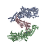
| Entry | Database: PDB / ID: 8w2p | ||||||
|---|---|---|---|---|---|---|---|
| Title | BsaXI-DNA complex I | ||||||
 Components Components |
| ||||||
 Keywords Keywords | DNA BINDING PROTEIN/DNA / restriction nuclease / restriction-modification systems / Type IIB / R-M complex / DNA BINDING PROTEIN / DNA BINDING PROTEIN-DNA complex | ||||||
| Function / homology |  Function and homology information Function and homology informationN-methyltransferase activity / DNA restriction-modification system / methylation / DNA binding Similarity search - Function | ||||||
| Biological species |   Geobacillus stearothermophilus (bacteria) Geobacillus stearothermophilus (bacteria) | ||||||
| Method | ELECTRON MICROSCOPY / single particle reconstruction / cryo EM / Resolution: 3.14 Å | ||||||
 Authors Authors | Shen, B.W. / Stoddard, B.L. / Xu, S. | ||||||
| Funding support |  United States, 1items United States, 1items
| ||||||
 Citation Citation | Journal: To Be Published Title: Structural and biochemical analysis of subunit assembly, DNA recognition and cleavage by a Type IIB restriction-modification enzyme: BsaXI Authors: Shen, B.W. / Heiter, D. / Xu, S. / Stoddard, B.L. | ||||||
| History |
|
- Structure visualization
| Structure viewer | Molecule: MolmilJmol/JSmol |
|---|
- Downloads & links
-Download
| PDBx/mmCIF format | 8w2p.cif.gz | 463 KB | Display | PDBx/mmCIF format |
|---|---|---|---|---|
| PDB format | pdb8w2p.ent.gz | 370.7 KB | Display | PDB format |
| PDBx/mmJSON format | 8w2p.json.gz | Tree view | PDBx/mmJSON format | |
| Others |  Other downloads Other downloads |
-Validation report
| Arichive directory | https://data.pdbj.org/pub/pdb/validation_reports/w2/8w2pftp://data.pdbj.org/pub/pdb/validation_reports/w2/8w2p | HTTPS FTP |
|---|
-Related structure data
| Related structure data | 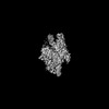 43754MC  8w2qC M: map data used to model this data C: citing same article ( |
|---|---|
| Similar structure data |

-Links
 PDBj
PDBj


- Assembly
| Deposited unit | 
|
|---|---|
| 1 |
|
-Components
-Protein , 2 types, 3 molecules ABC
| #1: Protein | Mass: 107195.234 Da / Num. of mol.: 2 Source method: isolated from a genetically manipulated source Source: (gene. exp.) Geobacillus stearothermophilus (bacteria)Strain: Cpw230 / Gene: B9L19_03590 / Production host: References: UniProt: A0AA91YUR9, site-specific DNA-methyltransferase (adenine-specific) #2: Protein | | Mass: 55038.824 Da / Num. of mol.: 1 Source method: isolated from a genetically manipulated source Source: (gene. exp.) Geobacillus stearothermophilus (bacteria)Strain: Cpw230 / Gene: GT3921_04730 / Production host: |
|---|
-DNA (41-MER) ... , 2 types, 2 molecules DE
| #3: DNA chain | Mass: 16083.387 Da / Num. of mol.: 1 / Source method: obtained synthetically / Source: (synth.) Geobacillus stearothermophilus (bacteria) |
|---|---|
| #4: DNA chain | Mass: 15963.281 Da / Num. of mol.: 1 / Source method: obtained synthetically / Source: (synth.) Geobacillus stearothermophilus (bacteria) |
-Non-polymers , 4 types, 8 molecules 


| #5: Chemical | ChemComp-SAM /  Mass: 398.437 Da / Num. of mol.: 1 / Source method: obtained synthetically / Formula: C15H22N6O5S / Feature type: SUBJECT OF INVESTIGATION Mass: 398.437 Da / Num. of mol.: 1 / Source method: obtained synthetically / Formula: C15H22N6O5S / Feature type: SUBJECT OF INVESTIGATION |
|---|---|
| #6: Chemical | ChemComp-CA /  Mass: 40.078 Da / Num. of mol.: 1 / Source method: obtained synthetically / Formula: Ca / Feature type: SUBJECT OF INVESTIGATION Mass: 40.078 Da / Num. of mol.: 1 / Source method: obtained synthetically / Formula: Ca / Feature type: SUBJECT OF INVESTIGATION |
| #7: Chemical | ChemComp-SAH /  Mass: 384.411 Da / Num. of mol.: 1 / Source method: obtained synthetically / Formula: C14H20N6O5S / Feature type: SUBJECT OF INVESTIGATION Mass: 384.411 Da / Num. of mol.: 1 / Source method: obtained synthetically / Formula: C14H20N6O5S / Feature type: SUBJECT OF INVESTIGATION |
| #8: Water | ChemComp-HOH / Mass: 18.015 Da / Num. of mol.: 5 / Source method: isolated from a natural source / Formula: H2O |
-Details
| Has ligand of interest | Y |
|---|---|
| Has protein modification | N |
-Experimental details
-Experiment
| Experiment | Method: ELECTRON MICROSCOPY |
|---|---|
| EM experiment | Aggregation state: PARTICLE / 3D reconstruction method: single particle reconstruction |
- Sample preparation
| Component | Name: Quaternary complex of RM, S and DNA complex / Type: COMPLEX / Entity ID: #1-#4 / Source: RECOMBINANT |
|---|---|
| Molecular weight | Value: 2.69 kDa/nm / Experimental value: NO |
| Source (natural) | Organism: Geobacillus stearothermophilus (bacteria) / Strain: Cpw230 |
| Source (recombinant) | Organism: |
| Buffer solution | pH: 8 / Details: 20 mM TrisHCl, 250 mM Nacl, 2 mM CaCl2 |
| Buffer component | Conc.: 20 mM / Name: TrisHCl |
| Specimen | Conc.: 0.3 mg/ml / Embedding applied: NO / Shadowing applied: NO / Staining applied: NO / Vitrification applied: YES / Details: Sample was monodisperse. |
| Specimen support | Grid material: COPPER / Grid mesh size: 300 divisions/in. / Grid type: Quantifoil R1.2/1.3 |
| Vitrification | Instrument: FEI VITROBOT MARK IV / Cryogen name: ETHANE / Humidity: 95 % / Chamber temperature: 278 K |
- Electron microscopy imaging
| Experimental equipment |  Model: Talos Arctica / Image courtesy: FEI Company /  Model: Titan Krios / Image courtesy: FEI Company | ||||||||||||||||||
|---|---|---|---|---|---|---|---|---|---|---|---|---|---|---|---|---|---|---|---|
| EM imaging | Alignment procedure: COMA FREE / C2 aperture diameter: 100 µm / Cryogen: NITROGEN / Electron source:
| ||||||||||||||||||
| Image recording | Average exposure time: 2 sec. / Electron dose: 40 e/Å2 / Detector mode: COUNTING
| ||||||||||||||||||
| EM imaging optics |
| ||||||||||||||||||
| Image scans |
|
 FIELD EMISSION GUN
- Processing
| EM software |
| |||||||||||||||||||||||||||||||||||||||||||||||||||||||
|---|---|---|---|---|---|---|---|---|---|---|---|---|---|---|---|---|---|---|---|---|---|---|---|---|---|---|---|---|---|---|---|---|---|---|---|---|---|---|---|---|---|---|---|---|---|---|---|---|---|---|---|---|---|---|---|---|
| CTF correction | Type: NONE | |||||||||||||||||||||||||||||||||||||||||||||||||||||||
| Symmetry | Point symmetry: C1 (asymmetric) | |||||||||||||||||||||||||||||||||||||||||||||||||||||||
| 3D reconstruction | Resolution: 3.14 Å / Resolution method: FSC 0.143 CUT-OFF / Num. of particles: 245111 / Algorithm: FOURIER SPACE / Num. of class averages: 2 / Symmetry type: POINT | |||||||||||||||||||||||||||||||||||||||||||||||||||||||
| Atomic model building | Protocol: BACKBONE TRACE / Space: REAL | |||||||||||||||||||||||||||||||||||||||||||||||||||||||
| Atomic model building | Details: manual built model in coot / Source name: Other / Type: experimental model | |||||||||||||||||||||||||||||||||||||||||||||||||||||||
| Refine LS restraints |
|
