 Movie
Movie Controller


+ Open data

- Basic information
| Entry | Database: PDB / ID: 8w12 | |||||||||
|---|---|---|---|---|---|---|---|---|---|---|
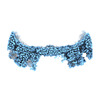
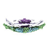
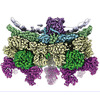
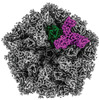
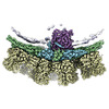
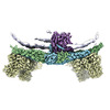
| Title | Cryo-EM structure of VP3-VP6 heterohexamer | |||||||||
 Components Components |
| |||||||||
 Keywords Keywords | VIRAL PROTEIN / Bluetongue virus / capsid protein / heterohexamer | |||||||||
| Function / homology | VP6 blue-tongue virus inner capsid protein / Orbivirus helicase VP6 / Inner layer core protein VP3, Orbivirus / Orbivirus VP3 (T2) protein / Inner layer core protein VP3, Reovirus / viral capsid / structural molecule activity / Core protein VP3 / VP6 Function and homology information Function and homology information | |||||||||
| Biological species |  Bluetongue virus Bluetongue virus | |||||||||
| Method | ELECTRON MICROSCOPY / single particle reconstruction / cryo EM / Resolution: 3.5 Å | |||||||||
 Authors Authors | Xia, X. / Sung, P.Y. / Martynowycz, M.W. / Gonen, T. / Roy, P. / Zhou, Z.H. | |||||||||
| Funding support |  United States, United States,  United Kingdom, 2items United Kingdom, 2items
| |||||||||
 Citation Citation | Journal: Cell / Year: 2024 Title: RNA genome packaging and capsid assembly of bluetongue virus visualized in host cells. Authors: Xian Xia / Po-Yu Sung / Michael W Martynowycz / Tamir Gonen / Polly Roy / Z Hong Zhou / Abstract: Unlike those of double-stranded DNA (dsDNA), single-stranded DNA (ssDNA), and ssRNA viruses, the mechanism of genome packaging of dsRNA viruses is poorly understood. Here, we combined the techniques ...Unlike those of double-stranded DNA (dsDNA), single-stranded DNA (ssDNA), and ssRNA viruses, the mechanism of genome packaging of dsRNA viruses is poorly understood. Here, we combined the techniques of high-resolution cryoelectron microscopy (cryo-EM), cellular cryoelectron tomography (cryo-ET), and structure-guided mutagenesis to investigate genome packaging and capsid assembly of bluetongue virus (BTV), a member of the Reoviridae family of dsRNA viruses. A total of eleven assembly states of BTV capsid were captured, with resolutions up to 2.8 Å, with most visualized in the host cytoplasm. ATPase VP6 was found underneath the vertices of capsid shell protein VP3 as an RNA-harboring pentamer, facilitating RNA packaging. RNA packaging expands the VP3 shell, which then engages middle- and outer-layer proteins to generate infectious virions. These revealed "duality" characteristics of the BTV assembly mechanism reconcile previous contradictory co-assembly and core-filling models and provide insights into the mysterious RNA packaging and capsid assembly of Reoviridae members and beyond. | |||||||||
| History |
|
- Structure visualization
| Structure viewer | Molecule: MolmilJmol/JSmol |
|---|
- Downloads & links
-Download
| PDBx/mmCIF format | 8w12.cif.gz | 646.7 KB | Display | PDBx/mmCIF format |
|---|---|---|---|---|
| PDB format | pdb8w12.ent.gz | 523.2 KB | Display | PDB format |
| PDBx/mmJSON format | 8w12.json.gz | Tree view | PDBx/mmJSON format | |
| Others |  Other downloads Other downloads |
-Validation report
| Arichive directory | https://data.pdbj.org/pub/pdb/validation_reports/w1/8w12ftp://data.pdbj.org/pub/pdb/validation_reports/w1/8w12 | HTTPS FTP |
|---|
-Related structure data
| Related structure data | 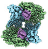 43714MC  8w19C  8w1cC  8w1iC  8w1oC  8w1rC  8w1sC M: map data used to model this data C: citing same article ( |
|---|---|
| Similar structure data |


-Links
 PDBj
PDBj
- Assembly
| Deposited unit | 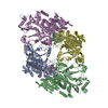
|
|---|---|
| 1 |
|
-Components
| #1: Protein | Mass: 105613.969 Da / Num. of mol.: 4 Source method: isolated from a genetically manipulated source Source: (gene. exp.) Bluetongue virus (serotype 1 / isolate South Africa)Strain: BTV1 / Gene: VP3 / Plasmid: pFastBac1 / Cell line (production host): Sf9 / Production host:   Spodoptera frugiperda (fall armyworm) / References: UniProt: Q1AE73 Spodoptera frugiperda (fall armyworm) / References: UniProt: Q1AE73#2: Protein | Mass: 22565.500 Da / Num. of mol.: 2 Source method: isolated from a genetically manipulated source Source: (gene. exp.) Bluetongue virus (serotype 1 / isolate South Africa)Strain: BTV1 / Plasmid: pFastBac1 / Cell line (production host): Sf9 / Production host: Spodoptera frugiperda (fall armyworm) / References: UniProt: Q91HQ0 |
|---|
-Experimental details
-Experiment
| Experiment | Method: ELECTRON MICROSCOPY |
|---|---|
| EM experiment | Aggregation state: PARTICLE / 3D reconstruction method: single particle reconstruction |
- Sample preparation
| Component | Name: Bluetongue virus (serotype 1 / isolate South Africa) / Type: VIRUS / Entity ID: all / Source: RECOMBINANT | ||||||||||||||||||||
|---|---|---|---|---|---|---|---|---|---|---|---|---|---|---|---|---|---|---|---|---|---|
| Molecular weight | Value: 0.46 MDa / Experimental value: YES | ||||||||||||||||||||
| Source (natural) | Organism: Bluetongue virus (serotype 1 / isolate South Africa) | ||||||||||||||||||||
| Source (recombinant) | Organism: Spodoptera frugiperda (fall armyworm) / Cell: sf9 / Plasmid: pFastBac1 | ||||||||||||||||||||
| Details of virus | Empty: YES / Enveloped: NO / Isolate: SPECIES / Type: VIRUS-LIKE PARTICLE | ||||||||||||||||||||
| Natural host | Organism: Ovis aries | ||||||||||||||||||||
| Buffer solution | pH: 7.5 | ||||||||||||||||||||
| Buffer component |
| ||||||||||||||||||||
| Specimen | Conc.: 1 mg/ml / Embedding applied: NO / Shadowing applied: NO / Staining applied: NO / Vitrification applied: YES Details: Recombinated VP3-VP6 complex purified from insect cell | ||||||||||||||||||||
| Specimen support | Grid material: COPPER / Grid mesh size: 200 divisions/in. / Grid type: Quantifoil R2/1 | ||||||||||||||||||||
| Vitrification | Instrument: FEI VITROBOT MARK IV / Cryogen name: ETHANE / Humidity: 100 % / Chamber temperature: 281.15 K |
- Electron microscopy imaging
| Experimental equipment |  Model: Titan Krios / Image courtesy: FEI Company |
|---|---|
| Microscopy | Model: FEI TITAN KRIOS |
| Electron gun | Electron source:  FIELD EMISSION GUN / Accelerating voltage: 300 kV / Illumination mode: FLOOD BEAM FIELD EMISSION GUN / Accelerating voltage: 300 kV / Illumination mode: FLOOD BEAM |
| Electron lens | Mode: BRIGHT FIELD / Nominal magnification: 81000 X / Nominal defocus max: 2600 nm / Nominal defocus min: 1800 nm / Cs: 2.7 mm / C2 aperture diameter: 50 µm / Alignment procedure: COMA FREE |
| Specimen holder | Cryogen: NITROGEN / Specimen holder model: FEI TITAN KRIOS AUTOGRID HOLDER |
| Image recording | Average exposure time: 2 sec. / Electron dose: 50 e/Å2 / Film or detector model: GATAN K3 BIOQUANTUM (6k x 4k) / Num. of grids imaged: 1 / Num. of real images: 2843 |
| EM imaging optics | Energyfilter name: GIF Bioquantum / Energyfilter slit width: 20 eV |
- Processing
| EM software |
| ||||||||||||||||||||||||||||||||||||
|---|---|---|---|---|---|---|---|---|---|---|---|---|---|---|---|---|---|---|---|---|---|---|---|---|---|---|---|---|---|---|---|---|---|---|---|---|---|
| CTF correction | Type: PHASE FLIPPING ONLY | ||||||||||||||||||||||||||||||||||||
| Particle selection | Num. of particles selected: 1451665 | ||||||||||||||||||||||||||||||||||||
| Symmetry | Point symmetry: C2 (2 fold cyclic) | ||||||||||||||||||||||||||||||||||||
| 3D reconstruction | Resolution: 3.5 Å / Resolution method: FSC 0.143 CUT-OFF / Num. of particles: 138115 / Num. of class averages: 1 / Symmetry type: POINT | ||||||||||||||||||||||||||||||||||||
| Atomic model building | B value: 107 / Protocol: AB INITIO MODEL / Space: REAL | ||||||||||||||||||||||||||||||||||||
| Refine LS restraints |
|
