 Movie
Movie Controller


+ Open data

- Basic information
| Entry | Database: PDB / ID: 8uxn | ||||||||||||||||||
|---|---|---|---|---|---|---|---|---|---|---|---|---|---|---|---|---|---|---|---|
| Title | Caulobacter crescentus FljM flagellar filament (symmetrized) | ||||||||||||||||||
 Components Components | Flagellin FljM | ||||||||||||||||||
 Keywords Keywords | STRUCTURAL PROTEIN / flagellin / flagellar filament | ||||||||||||||||||
| Function / homology | Flagellin, C-terminal domain / Bacterial flagellin C-terminal helical region / Flagellin / Flagellin, N-terminal domain / Bacterial flagellin N-terminal helical region / bacterial-type flagellum / structural molecule activity / extracellular region / Flagellin FljM Function and homology information Function and homology information | ||||||||||||||||||
| Biological species |  Caulobacter vibrioides (bacteria) Caulobacter vibrioides (bacteria) | ||||||||||||||||||
| Method | ELECTRON MICROSCOPY / helical reconstruction / cryo EM / Resolution: 2.11 Å | ||||||||||||||||||
 Authors Authors | Sanchez, J.C. / Montemayor, E.J. / Ploscariu, N.T. / Parrell, D. / Baumgardt, J.K. / Yang, J.E. / Sibert, B. / Cai, K. / Wright, E.R. | ||||||||||||||||||
| Funding support |  United States, 5items United States, 5items
| ||||||||||||||||||
 Citation Citation | Journal: To Be Published Title: Direct evidence for multi-flagellin filament stabilization via atomic-level architecture of Caulobacter crescentus flagellar filaments Authors: Sanchez, J.C. / Montemayor, E.J. / Ploscariu, N.T. / Parrell, D. / Baumgardt, J.K. / Yang, J.E. / Sibert, B. / Cai, K. / Wright, E.R. | ||||||||||||||||||
| History |
|
- Structure visualization
| Structure viewer | Molecule: MolmilJmol/JSmol |
|---|
- Downloads & links
-Download
| PDBx/mmCIF format | 8uxn.cif.gz | 1.9 MB | Display | PDBx/mmCIF format |
|---|---|---|---|---|
| PDB format | pdb8uxn.ent.gz | 1.6 MB | Display | PDB format |
| PDBx/mmJSON format | 8uxn.json.gz | Tree view | PDBx/mmJSON format | |
| Others |  Other downloads Other downloads |
-Validation report
| Summary document | 8uxn_validation.pdf.gz | 1.3 MB | Display | wwPDB validaton report |
|---|---|---|---|---|
| Full document | 8uxn_full_validation.pdf.gz | 1.3 MB | Display | |
| Data in XML | 8uxn_validation.xml.gz | 233.3 KB | Display | |
| Data in CIF | 8uxn_validation.cif.gz | 347.6 KB | Display | |
| Arichive directory | https://data.pdbj.org/pub/pdb/validation_reports/ux/8uxnftp://data.pdbj.org/pub/pdb/validation_reports/ux/8uxn | HTTPS FTP |
-Related structure data
| Related structure data | 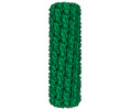 42770MC M: map data used to model this data C: citing same article ( |
|---|---|
| Similar structure data |
-Links
 PDBj
PDBj- Assembly
| Deposited unit | 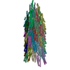
|
|---|---|
| 1 |
|
-Components
| #1: Protein | Mass: 27950.240 Da / Num. of mol.: 44 / Source method: isolated from a natural source / Source: (natural) Caulobacter vibrioides (bacteria) / Strain: NA1000 / References: UniProt: O52529 |
|---|
-Experimental details
-Experiment
| Experiment | Method: ELECTRON MICROSCOPY |
|---|---|
| EM experiment | Aggregation state: FILAMENT / 3D reconstruction method: helical reconstruction |
- Sample preparation
| Component | Name: FljM flagellar filament (symmetrized) / Type: COMPLEX / Entity ID: all / Source: NATURAL |
|---|---|
| Molecular weight | Value: 0.28 kDa/nm / Experimental value: NO |
| Source (natural) | Organism: Caulobacter vibrioides (bacteria) / Strain: NA1000 |
| Buffer solution | pH: 7.4 |
| Buffer component | Name: phosphate buffered saline |
| Specimen | Embedding applied: NO / Shadowing applied: NO / Staining applied: NO / Vitrification applied: YES |
| Specimen support | Grid material: COPPER / Grid mesh size: 200 divisions/in. / Grid type: Quantifoil R2/1 |
| Vitrification | Instrument: FEI VITROBOT MARK IV / Cryogen name: ETHANE / Humidity: 95 % / Chamber temperature: 293 K |
- Electron microscopy imaging
| Experimental equipment |  Model: Titan Krios / Image courtesy: FEI Company |
|---|---|
| Microscopy | Model: TFS KRIOS |
| Electron gun | Electron source:  FIELD EMISSION GUN / Accelerating voltage: 300 kV / Illumination mode: FLOOD BEAM FIELD EMISSION GUN / Accelerating voltage: 300 kV / Illumination mode: FLOOD BEAM |
| Electron lens | Mode: BRIGHT FIELD / Nominal magnification: 105000 X / Nominal defocus max: 2500 nm / Nominal defocus min: 500 nm / C2 aperture diameter: 70 µm |
| Specimen holder | Specimen holder model: FEI TITAN KRIOS AUTOGRID HOLDER |
| Image recording | Electron dose: 45 e/Å2 / Film or detector model: GATAN K3 BIOQUANTUM (6k x 4k) |
| EM imaging optics | Energyfilter name: GIF Bioquantum / Energyfilter slit width: 20 eV |
- Processing
| Software | Name: PHENIX / Version: 1.20.1_4487: / Classification: refinement | |||||||||||||||||||||||||||||||||||||||||||||||||||||||
|---|---|---|---|---|---|---|---|---|---|---|---|---|---|---|---|---|---|---|---|---|---|---|---|---|---|---|---|---|---|---|---|---|---|---|---|---|---|---|---|---|---|---|---|---|---|---|---|---|---|---|---|---|---|---|---|---|
| EM software |
| |||||||||||||||||||||||||||||||||||||||||||||||||||||||
| CTF correction | Type: PHASE FLIPPING AND AMPLITUDE CORRECTION | |||||||||||||||||||||||||||||||||||||||||||||||||||||||
| Helical symmerty | Angular rotation/subunit: 65.93 ° / Axial rise/subunit: 4.73 Å / Axial symmetry: C1 | |||||||||||||||||||||||||||||||||||||||||||||||||||||||
| 3D reconstruction | Resolution: 2.11 Å / Resolution method: FSC 0.143 CUT-OFF / Num. of particles: 248115 / Symmetry type: HELICAL | |||||||||||||||||||||||||||||||||||||||||||||||||||||||
| Atomic model building | B value: 67 / Space: REAL | |||||||||||||||||||||||||||||||||||||||||||||||||||||||
| Atomic model building | Accession code: O52529 / Source name: AlphaFold | |||||||||||||||||||||||||||||||||||||||||||||||||||||||
| Refine LS restraints |
|
