 Movie
Movie Controller

[English] 日本語
 Yorodumi
- PDB-8uhe: Structure of the far-red light-absorbing allophycocyanin core exp... -
+ Open data

- Basic information
| Entry | Database: PDB / ID: 8uhe | ||||||||||||
|---|---|---|---|---|---|---|---|---|---|---|---|---|---|
| Title | Structure of the far-red light-absorbing allophycocyanin core expressed during FaRLiP | ||||||||||||
 Components Components |
| ||||||||||||
 Keywords Keywords | PHOTOSYNTHESIS / light-harvesting / allophycocyanin / phycocyanobilin | ||||||||||||
| Function / homology |  Function and homology information Function and homology informationphycobilisome / plasma membrane-derived thylakoid membrane / photosynthesis / lyase activity Similarity search - Function | ||||||||||||
| Biological species |  Synechococcus sp. PCC 7335 (bacteria) Synechococcus sp. PCC 7335 (bacteria) | ||||||||||||
| Method | ELECTRON MICROSCOPY / single particle reconstruction / cryo EM / Resolution: 2.78 Å | ||||||||||||
 Authors Authors | Gisriel, C.J. / Bryant, D.A. / Brudvig, G.W. / Shen, G. | ||||||||||||
| Funding support |  United States, 3items United States, 3items
| ||||||||||||
 Citation Citation | Journal: J Biol Chem / Year: 2024 Title: Structure of the antenna complex expressed during far-red light photoacclimation in Synechococcus sp. PCC 7335. Authors: Christopher J Gisriel / Gaozhong Shen / Gary W Brudvig / Donald A Bryant / Abstract: Far-red light photoacclimation, or FaRLiP, is a facultative response exhibited by some cyanobacteria that allows them to absorb and utilize lower energy light (700-800 nm) than the wavelengths ...Far-red light photoacclimation, or FaRLiP, is a facultative response exhibited by some cyanobacteria that allows them to absorb and utilize lower energy light (700-800 nm) than the wavelengths typically used for oxygenic photosynthesis (400-700 nm). During this process, three essential components of the photosynthetic apparatus are altered: photosystem I, photosystem II, and the phycobilisome. In all three cases, at least some of the chromophores found in these pigment-protein complexes are replaced by chromophores that have red-shifted absorbance relative to the analogous complexes produced in visible light. Recent structural and spectroscopic studies have elucidated important features of the two photosystems when altered to absorb and utilize far-red light, but much less is understood about the modified phycobiliproteins made during FaRLiP. We used single-particle, cryo-EM to determine the molecular structure of a phycobiliprotein core complex comprising allophycocyanin variants that absorb far-red light during FaRLiP in the marine cyanobacterium Synechococcus sp. PCC 7335. The structure reveals the arrangement of the numerous red-shifted allophycocyanin variants and the probable locations of the chromophores that serve as the terminal emitters in this complex. It also suggests how energy is transferred to the photosystem II complexes produced during FaRLiP. The structure additionally allows comparisons with other previously studied allophycocyanins to gain insights into how phycocyanobilin chromophores can be tuned to absorb far-red light. These studies provide new insights into how far-red light is harvested and utilized during FaRLiP, a widespread cyanobacterial photoacclimation mechanism. | ||||||||||||
| History |
|
- Structure visualization
| Structure viewer | Molecule: MolmilJmol/JSmol |
|---|
- Downloads & links
-Download
| PDBx/mmCIF format | 8uhe.cif.gz | 658.8 KB | Display | PDBx/mmCIF format |
|---|---|---|---|---|
| PDB format | pdb8uhe.ent.gz | 542.2 KB | Display | PDB format |
| PDBx/mmJSON format | 8uhe.json.gz | Tree view | PDBx/mmJSON format | |
| Others |  Other downloads Other downloads |
-Validation report
| Arichive directory | https://data.pdbj.org/pub/pdb/validation_reports/uh/8uheftp://data.pdbj.org/pub/pdb/validation_reports/uh/8uhe | HTTPS FTP |
|---|
-Related structure data
| Related structure data |  42278MC 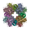 8uhiC M: map data used to model this data C: citing same article ( |
|---|---|
| Similar structure data |

-Links
 PDBj
PDBj- Assembly
| Deposited unit | 
|
|---|---|
| 1 |
|
-Components
-Protein , 7 types, 19 molecules AEGMOQBDFHLNPRCIJKS
| #1: Protein | Mass: 17837.252 Da / Num. of mol.: 6 / Source method: isolated from a natural source / Source: (natural) Synechococcus sp. PCC 7335 (bacteria) / References: UniProt: B4WKI9#2: Protein | Mass: 17501.822 Da / Num. of mol.: 8 / Source method: isolated from a natural source / Source: (natural) Synechococcus sp. PCC 7335 (bacteria) / References: UniProt: B4WKI8#3: Protein | | Mass: 20339.404 Da / Num. of mol.: 1 / Source method: isolated from a natural source / Source: (natural) Synechococcus sp. PCC 7335 (bacteria) / References: UniProt: B4WKI4#4: Protein | | Mass: 17891.504 Da / Num. of mol.: 1 / Source method: isolated from a natural source / Source: (natural) Synechococcus sp. PCC 7335 (bacteria) / References: UniProt: B4WKI7#5: Protein | | Mass: 18950.438 Da / Num. of mol.: 1 / Source method: isolated from a natural source / Source: (natural) Synechococcus sp. PCC 7335 (bacteria) / References: UniProt: B4WIH2#6: Protein | | Mass: 87252.891 Da / Num. of mol.: 1 / Source method: isolated from a natural source / Source: (natural) Synechococcus sp. PCC 7335 (bacteria) / References: UniProt: B4WKI6#7: Protein | | Mass: 7804.070 Da / Num. of mol.: 1 / Source method: isolated from a natural source / Source: (natural) Synechococcus sp. PCC 7335 (bacteria) / References: UniProt: B4WI75 |
|---|
-Non-polymers , 3 types, 19 molecules 

| #8: Chemical | ChemComp-CYC /  Mass: 588.694 Da / Num. of mol.: 16 / Source method: obtained synthetically / Formula: C33H40N4O6 Mass: 588.694 Da / Num. of mol.: 16 / Source method: obtained synthetically / Formula: C33H40N4O6#9: Chemical |  Mass: 586.678 Da / Num. of mol.: 2 / Source method: obtained synthetically / Formula: C33H38N4O6 / Feature type: SUBJECT OF INVESTIGATION Mass: 586.678 Da / Num. of mol.: 2 / Source method: obtained synthetically / Formula: C33H38N4O6 / Feature type: SUBJECT OF INVESTIGATION#10: Chemical | ChemComp-CL / |  Mass: 35.453 Da / Num. of mol.: 1 / Source method: obtained synthetically / Formula: Cl Mass: 35.453 Da / Num. of mol.: 1 / Source method: obtained synthetically / Formula: Cl |
|---|
-Details
| Has ligand of interest | Y |
|---|
-Experimental details
-Experiment
| Experiment | Method: ELECTRON MICROSCOPY |
|---|---|
| EM experiment | Aggregation state: PARTICLE / 3D reconstruction method: single particle reconstruction |
- Sample preparation
| Component | Name: Far-red light-absorbing allophycocyanin core / Type: COMPLEX / Entity ID: #1-#7 / Source: NATURAL |
|---|---|
| Source (natural) | Organism: Synechococcus sp. PCC 7335 (bacteria) |
| Buffer solution | pH: 7 |
| Specimen | Embedding applied: NO / Shadowing applied: NO / Staining applied: NO / Vitrification applied: YES |
| Vitrification | Cryogen name: ETHANE |
- Electron microscopy imaging
| Experimental equipment |  Model: Titan Krios / Image courtesy: FEI Company |
|---|---|
| Microscopy | Model: FEI TITAN KRIOS |
| Electron gun | Electron source:  FIELD EMISSION GUN / Accelerating voltage: 300 kV / Illumination mode: FLOOD BEAM FIELD EMISSION GUN / Accelerating voltage: 300 kV / Illumination mode: FLOOD BEAM |
| Electron lens | Mode: BRIGHT FIELD / Nominal defocus max: 2200 nm / Nominal defocus min: 800 nm |
| Image recording | Electron dose: 50 e/Å2 / Film or detector model: GATAN K3 (6k x 4k) |
- Processing
| CTF correction | Type: PHASE FLIPPING AND AMPLITUDE CORRECTION |
|---|---|
| 3D reconstruction | Resolution: 2.78 Å / Resolution method: FSC 0.143 CUT-OFF / Num. of particles: 113162 / Symmetry type: POINT |
