National Institutes of Health/National Institute of General Medical Sciences (NIH/NIGMS)
R01-GM127593
米国
引用
ジャーナル: Nat Struct Mol Biol / 年: 2024 タイトル: The LexA-RecA* structure reveals a cryptic lock-and-key mechanism for SOS activation. 著者: Michael B Cory / Allen Li / Christina M Hurley / Peter J Carman / Ruth A Pumroy / Zachary M Hostetler / Ryann M Perez / Yarra Venkatesh / Xinning Li / Kushol Gupta / E James Petersson / Rahul M Kohli / 要旨: The bacterial SOS response plays a key role in adaptation to DNA damage, including genomic stress caused by antibiotics. SOS induction begins when activated RecA*, an oligomeric nucleoprotein ...The bacterial SOS response plays a key role in adaptation to DNA damage, including genomic stress caused by antibiotics. SOS induction begins when activated RecA*, an oligomeric nucleoprotein filament that forms on single-stranded DNA, binds to and stimulates autoproteolysis of the repressor LexA. Here, we present the structure of the complete Escherichia coli SOS signal complex, constituting full-length LexA bound to RecA*. We uncover an extensive interface unexpectedly including the LexA DNA-binding domain, providing a new molecular rationale for ordered SOS gene induction. We further find that the interface involves three RecA subunits, with a single residue in the central engaged subunit acting as a molecular key, inserting into an allosteric binding pocket to induce LexA cleavage. Given the pro-mutagenic nature of SOS activation, our structural and mechanistic insights provide a foundation for developing new therapeutics to slow the evolution of antibiotic resistance.
#241 - 2020年1月 20年の分子を振り返って (Twenty Years of Molecules) 類似性 (1)
-
集合体
登録構造単位
A: Protein RecA B: Protein RecA C: Protein RecA D: Protein RecA E: Protein RecA F: Protein RecA G: Protein RecA H: Protein RecA I: LexA repressor J: LexA repressor L: DNA (27-MER) ヘテロ分子
モード: BRIGHT FIELD / 倍率(公称値): 64000 X / 最大 デフォーカス(公称値): 2000 nm / 最小 デフォーカス(公称値): 500 nm / Cs: 2.7 mm / C2レンズ絞り径: 100 µm
試料ホルダ
試料ホルダーモデル: FEI TITAN KRIOS AUTOGRID HOLDER
撮影
平均露光時間: 5.35 sec. / 電子線照射量: 46.2 e/Å2 / フィルム・検出器のモデル: GATAN K3 (6k x 4k) / 撮影したグリッド数: 1 / 実像数: 2748 / 詳細: 40 frames collected per movie
-
解析
EMソフトウェア
ID
名称
バージョン
カテゴリ
詳細
1
cryoSPARC
4.0.3
粒子像選択
FilamentTracer
2
EPU
画像取得
4
cryoSPARC
4.0.3
CTF補正
PatchCTFEstimation
7
UCSF ChimeraX
1.3
モデルフィッティング
9
PHENIX
1.20.1
モデル精密化
10
ISOLDE
1.4
モデル精密化
13
cryoSPARC
4.0.3
分類
3DClassification
14
cryoSPARC
4.0.3
3次元再構成
LocalRefinement
CTF補正
タイプ: PHASE FLIPPING AND AMPLITUDE CORRECTION
らせん対称
ID
Image processing-ID
回転角度/サブユニット (°)
軸方向距離/サブユニット (Å)
らせん対称軸の対称性
1
1
59.2
16.23
C1
2
1
59.2
16.23
C1
3
1
59.2
16.23
C1
4
1
59.2
16.23
C1
粒子像の選択
選択した粒子像数: 3423408 詳細: cryoSPARC automated Filament Tracer using filament diameter of 100A, 17A separation between segments. Minimum and maximum filament diameters of 90A, 150A respectively. Inspect Picks to filter ...詳細: cryoSPARC automated Filament Tracer using filament diameter of 100A, 17A separation between segments. Minimum and maximum filament diameters of 90A, 150A respectively. Inspect Picks to filter particles with high curvature or sinuosity Local power > 567.940 Local power < 5711.103 Curvature (1/A) < 0.005970 Sinuosity < 1.393924 Low pass filtered particles near micrograph edge.
 ムービー
ムービー コントローラー


 データを開く
 基本情報
基本情報 要素
要素 キーワード
キーワード 機能・相同性情報
 データ登録者
 米国, 1件
  引用
引用 構造の表示
構造の表示 ダウンロードとリンク
 その他のダウンロード
 PDBj
PDBj


 集合体
集合体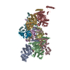

 分子量: 523.247 Da / 分子数: 8 / 由来タイプ: 合成 / 式: C10H16N5O12P3S
 分子量: 24.305 Da / 分子数: 8 / 由来タイプ: 合成 / 式: Mg
 試料調製
試料調製 電子顕微鏡撮影
 FIELD EMISSION GUN / 加速電圧: 300 kV / 照射モード: SPOT SCAN
 解析
解析