 Movie
Movie Controller


+ Open data

- Basic information
| Entry | Database: PDB / ID: 8t3t | ||||||
|---|---|---|---|---|---|---|---|
| Title | Structure of Bre1-nucleosome complex - state3 | ||||||
 Components Components |
| ||||||
 Keywords Keywords | DNA BINDING PROTEIN/Transferase/DNA / H2B ubiquitin E3 ligase / dimer / nucleosome acidic patch binding protein / DNA BINDING PROTEIN-Transferase-DNA complex | ||||||
| Function / homology |  Function and homology information Function and homology informationHULC complex / meiotic DNA double-strand break formation / telomere maintenance via recombination / mitotic intra-S DNA damage checkpoint signaling / regulation of DNA-templated DNA replication initiation / DNA replication origin binding / subtelomeric heterochromatin formation / mitotic G1 DNA damage checkpoint signaling / RING-type E3 ubiquitin transferase / double-strand break repair via homologous recombination ...HULC complex / meiotic DNA double-strand break formation / telomere maintenance via recombination / mitotic intra-S DNA damage checkpoint signaling / regulation of DNA-templated DNA replication initiation / DNA replication origin binding / subtelomeric heterochromatin formation / mitotic G1 DNA damage checkpoint signaling / RING-type E3 ubiquitin transferase / double-strand break repair via homologous recombination / structural constituent of chromatin / ubiquitin protein ligase activity / nucleosome / heterochromatin formation / nucleosome assembly / scaffold protein binding / transcription by RNA polymerase II / chromosome, telomeric region / protein ubiquitination / protein heterodimerization activity / chromatin / DNA binding / zinc ion binding / nucleoplasm / identical protein binding / nucleus Similarity search - Function | ||||||
| Biological species |  synthetic construct (others) | ||||||
| Method | ELECTRON MICROSCOPY / single particle reconstruction / cryo EM / Resolution: 3.21 Å | ||||||
 Authors Authors | Zhao, F. / Hicks, C.W. / Wolberger, C. | ||||||
| Funding support |  United States, 1items United States, 1items
| ||||||
 Citation Citation | Journal: Nat Struct Mol Biol / Year: 2023 Title: Mechanism of histone H2B monoubiquitination by Bre1. Authors: Fan Zhao / Chad W Hicks / Cynthia Wolberger / Abstract: Monoubiquitination of histone H2B-K120/123 plays several roles in regulating transcription, DNA replication and the DNA damage response. The structure of a nucleosome in complex with the dimeric RING ...Monoubiquitination of histone H2B-K120/123 plays several roles in regulating transcription, DNA replication and the DNA damage response. The structure of a nucleosome in complex with the dimeric RING E3 ligase Bre1 reveals that one RING domain binds to the nucleosome acidic patch, where it can position the E2 ubiquitin conjugating enzyme Rad6, while the other RING domain contacts the DNA. Comparisons with H2A-specific E3 ligases suggest a general mechanism of tuning histone specificity via the non-E2-binding RING domain. | ||||||
| History |
|
- Structure visualization
| Structure viewer | Molecule: MolmilJmol/JSmol |
|---|
- Downloads & links
-Download
| PDBx/mmCIF format | 8t3t.cif.gz | 323.7 KB | Display | PDBx/mmCIF format |
|---|---|---|---|---|
| PDB format | pdb8t3t.ent.gz | 260.2 KB | Display | PDB format |
| PDBx/mmJSON format | 8t3t.json.gz | Tree view | PDBx/mmJSON format | |
| Others |  Other downloads Other downloads |
-Validation report
| Arichive directory | https://data.pdbj.org/pub/pdb/validation_reports/t3/8t3tftp://data.pdbj.org/pub/pdb/validation_reports/t3/8t3t | HTTPS FTP |
|---|
-Related structure data
| Related structure data |  41011MC  8t3wC  8t3yC M: map data used to model this data C: citing same article ( |
|---|---|
| Similar structure data |


-Links
 PDBj
PDBj


- Assembly
| Deposited unit | 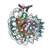
|
|---|---|
| 1 |
|
-Components
-Protein , 5 types, 10 molecules AEBFCGDHKL
| #1: Protein | Mass: 15303.930 Da / Num. of mol.: 2 Source method: isolated from a genetically manipulated source Source: (gene. exp.)  #2: Protein | Mass: 11263.231 Da / Num. of mol.: 2 Source method: isolated from a genetically manipulated source Source: (gene. exp.) #3: Protein | Mass: 13978.241 Da / Num. of mol.: 2 Source method: isolated from a genetically manipulated source Source: (gene. exp.) #4: Protein | Mass: 13524.752 Da / Num. of mol.: 2 Source method: isolated from a genetically manipulated source Source: (gene. exp.) #7: Protein | Mass: 12633.649 Da / Num. of mol.: 2 Source method: isolated from a genetically manipulated source Source: (gene. exp.) Gene: BRE1, YDL074C / Production host: References: UniProt: Q07457, RING-type E3 ubiquitin transferase |
|---|
-601 DNA strand ... , 2 types, 2 molecules IJ
| #5: DNA chain | Mass: 44825.559 Da / Num. of mol.: 1 / Source method: obtained synthetically / Source: (synth.) synthetic construct (others) |
|---|---|
| #6: DNA chain | Mass: 45305.852 Da / Num. of mol.: 1 / Source method: obtained synthetically / Source: (synth.) synthetic construct (others) |
-Non-polymers , 1 types, 4 molecules 
| #8: Chemical | ChemComp-ZN /  Mass: 65.409 Da / Num. of mol.: 4 / Source method: obtained synthetically / Formula: Zn / Feature type: SUBJECT OF INVESTIGATION Mass: 65.409 Da / Num. of mol.: 4 / Source method: obtained synthetically / Formula: Zn / Feature type: SUBJECT OF INVESTIGATION |
|---|
-Details
| Has ligand of interest | Y |
|---|
-Experimental details
-Experiment
| Experiment | Method: ELECTRON MICROSCOPY |
|---|---|
| EM experiment | Aggregation state: PARTICLE / 3D reconstruction method: single particle reconstruction |
- Sample preparation
| Component | Name: Bre1-nucleosome complex / Type: COMPLEX / Entity ID: #1-#7 / Source: RECOMBINANT |
|---|---|
| Source (natural) | Organism: |
| Source (recombinant) | Organism: |
| Buffer solution | pH: 7.5 / Details: 20 mM HEPES pH 7.5, 50 mM NaCl, 1 mM DTT |
| Specimen | Conc.: 1 mg/ml / Embedding applied: NO / Shadowing applied: NO / Staining applied: NO / Vitrification applied: YES / Details: Crosslinked with glutaraldehyde |
| Vitrification | Instrument: FEI VITROBOT MARK IV / Cryogen name: ETHANE / Humidity: 100 % / Chamber temperature: 277.15 K |
- Electron microscopy imaging
| Experimental equipment |  Model: Titan Krios / Image courtesy: FEI Company |
|---|---|
| Microscopy | Model: FEI TITAN KRIOS |
| Electron gun | Electron source:  FIELD EMISSION GUN / Accelerating voltage: 300 kV / Illumination mode: FLOOD BEAM FIELD EMISSION GUN / Accelerating voltage: 300 kV / Illumination mode: FLOOD BEAM |
| Electron lens | Mode: BRIGHT FIELD / Nominal defocus max: 2500 nm / Nominal defocus min: 1250 nm / Cs: 2.7 mm / C2 aperture diameter: 100 µm |
| Image recording | Electron dose: 50.02 e/Å2 / Film or detector model: GATAN K3 (6k x 4k) |
- Processing
| EM software |
| ||||||||||||||||||||||||
|---|---|---|---|---|---|---|---|---|---|---|---|---|---|---|---|---|---|---|---|---|---|---|---|---|---|
| CTF correction | Type: PHASE FLIPPING AND AMPLITUDE CORRECTION | ||||||||||||||||||||||||
| 3D reconstruction | Resolution: 3.21 Å / Resolution method: FSC 0.143 CUT-OFF / Num. of particles: 170468 / Symmetry type: POINT | ||||||||||||||||||||||||
| Atomic model building |
| ||||||||||||||||||||||||
| Refine LS restraints |
|
