 Movie
Movie Controller

[English] 日本語
 Yorodumi
- PDB-8si6: Cryo-EM structure of TRPM7 in MSP2N2 nanodisc in complex with ago... -
+ Open data

- Basic information
| Entry | Database: PDB / ID: 8si6 | ||||||||||||||||||
|---|---|---|---|---|---|---|---|---|---|---|---|---|---|---|---|---|---|---|---|
| Title | Cryo-EM structure of TRPM7 in MSP2N2 nanodisc in complex with agonist naltriben in closed state | ||||||||||||||||||
 Components Components | Transient receptor potential cation channel subfamily M member 7 | ||||||||||||||||||
 Keywords Keywords | MEMBRANE PROTEIN / transient receptor potential M family member 7 / TRP / channel / TRPM7 / TRP channels / magnesium channel / agonist / ligand / naltriben | ||||||||||||||||||
| Function / homology |  Function and homology information Function and homology informationcalcium-dependent cell-matrix adhesion / intracellular magnesium ion homeostasis / magnesium ion transmembrane transport / zinc ion transport / zinc ion transmembrane transporter activity / magnesium ion transmembrane transporter activity / TRP channels / actomyosin structure organization / myosin binding / necroptotic process ...calcium-dependent cell-matrix adhesion / intracellular magnesium ion homeostasis / magnesium ion transmembrane transport / zinc ion transport / zinc ion transmembrane transporter activity / magnesium ion transmembrane transporter activity / TRP channels / actomyosin structure organization / myosin binding / necroptotic process / ruffle / cytoplasmic vesicle membrane / calcium channel activity / calcium ion transport / kinase activity / actin binding / protein autophosphorylation / cytoplasmic vesicle / protein homotetramerization / non-specific serine/threonine protein kinase / protein kinase activity / protein serine kinase activity / protein serine/threonine kinase activity / ATP binding / metal ion binding / nucleus / plasma membrane Similarity search - Function | ||||||||||||||||||
| Biological species |  | ||||||||||||||||||
| Method | ELECTRON MICROSCOPY / single particle reconstruction / cryo EM / Resolution: 2.44 Å | ||||||||||||||||||
 Authors Authors | Nadezhdin, K.D. / Neuberger, A. / Sobolevsky, A.I. | ||||||||||||||||||
| Funding support |  United States, United States,  Germany, 5items Germany, 5items
| ||||||||||||||||||
 Citation Citation | Journal: Nat Commun / Year: 2023 Title: Structural mechanisms of TRPM7 activation and inhibition. Authors: Kirill D Nadezhdin / Leonor Correia / Chamali Narangoda / Dhilon S Patel / Arthur Neuberger / Thomas Gudermann / Maria G Kurnikova / Vladimir Chubanov / Alexander I Sobolevsky / Abstract: The transient receptor potential channel TRPM7 is a master regulator of the organismal balance of divalent cations that plays an essential role in embryonic development, immune responses, cell ...The transient receptor potential channel TRPM7 is a master regulator of the organismal balance of divalent cations that plays an essential role in embryonic development, immune responses, cell mobility, proliferation, and differentiation. TRPM7 is implicated in neuronal and cardiovascular disorders, tumor progression and has emerged as a new drug target. Here we use cryo-EM, functional analysis, and molecular dynamics simulations to uncover two distinct structural mechanisms of TRPM7 activation by a gain-of-function mutation and by the agonist naltriben, which show different conformational dynamics and domain involvement. We identify a binding site for highly potent and selective inhibitors and show that they act by stabilizing the TRPM7 closed state. The discovered structural mechanisms provide foundations for understanding the molecular basis of TRPM7 channelopathies and drug development. | ||||||||||||||||||
| History |
|
- Structure visualization
| Structure viewer | Molecule: MolmilJmol/JSmol |
|---|
- Downloads & links
-Download
| PDBx/mmCIF format | 8si6.cif.gz | 1.6 MB | Display | PDBx/mmCIF format |
|---|---|---|---|---|
| PDB format | pdb8si6.ent.gz | 1.4 MB | Display | PDB format |
| PDBx/mmJSON format | 8si6.json.gz | Tree view | PDBx/mmJSON format | |
| Others |  Other downloads Other downloads |
-Validation report
| Summary document | 8si6_validation.pdf.gz | 2.9 MB | Display | wwPDB validaton report |
|---|---|---|---|---|
| Full document | 8si6_full_validation.pdf.gz | 3 MB | Display | |
| Data in XML | 8si6_validation.xml.gz | 146.3 KB | Display | |
| Data in CIF | 8si6_validation.cif.gz | 207.4 KB | Display | |
| Arichive directory | https://data.pdbj.org/pub/pdb/validation_reports/si/8si6ftp://data.pdbj.org/pub/pdb/validation_reports/si/8si6 | HTTPS FTP |
-Related structure data
| Related structure data |  40500MC  8si2C  8si3C  8si4C  8si5C  8si7C  8si8C  8siaC  8sibC C: citing same article ( M: map data used to model this data |
|---|---|
| Similar structure data |


-Links
 PDBj
PDBj


- Assembly
| Deposited unit | 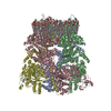
|
|---|---|
| 1 |
|
-Components
-Protein , 1 types, 4 molecules ABCD
| #1: Protein | Mass: 146888.875 Da / Num. of mol.: 4 Source method: isolated from a genetically manipulated source Source: (gene. exp.)  Homo sapiens (human) Homo sapiens (human)References: UniProt: Q923J1, non-specific serine/threonine protein kinase |
|---|
-Non-polymers , 5 types, 65 molecules 


| #2: Chemical | ChemComp-POV / (  Mass: 760.076 Da / Num. of mol.: 52 / Source method: obtained synthetically / Formula: C42H82NO8P / Comment: phospholipid*YM Mass: 760.076 Da / Num. of mol.: 52 / Source method: obtained synthetically / Formula: C42H82NO8P / Comment: phospholipid*YM#3: Chemical | ChemComp-CLR /  Mass: 386.654 Da / Num. of mol.: 4 / Source method: obtained synthetically / Formula: C27H46O Mass: 386.654 Da / Num. of mol.: 4 / Source method: obtained synthetically / Formula: C27H46O#4: Chemical | ChemComp-DU0 /  Mass: 516.752 Da / Num. of mol.: 4 / Source method: obtained synthetically / Formula: C32H52O5 Mass: 516.752 Da / Num. of mol.: 4 / Source method: obtained synthetically / Formula: C32H52O5#5: Chemical | ChemComp-ZY8 / (  Mass: 415.481 Da / Num. of mol.: 4 / Source method: obtained synthetically / Formula: C26H25NO4 / Feature type: SUBJECT OF INVESTIGATION Mass: 415.481 Da / Num. of mol.: 4 / Source method: obtained synthetically / Formula: C26H25NO4 / Feature type: SUBJECT OF INVESTIGATION#6: Chemical | ChemComp-CA / |  Mass: 40.078 Da / Num. of mol.: 1 / Source method: obtained synthetically / Formula: Ca Mass: 40.078 Da / Num. of mol.: 1 / Source method: obtained synthetically / Formula: Ca |
|---|
-Details
| Has ligand of interest | Y |
|---|---|
| Has protein modification | Y |
-Experimental details
-Experiment
| Experiment | Method: ELECTRON MICROSCOPY |
|---|---|
| EM experiment | Aggregation state: PARTICLE / 3D reconstruction method: single particle reconstruction |
- Sample preparation
| Component | Name: sample 1 / Type: COMPLEX / Entity ID: #1 / Source: RECOMBINANT | ||||||||||||||||||||||||||||||
|---|---|---|---|---|---|---|---|---|---|---|---|---|---|---|---|---|---|---|---|---|---|---|---|---|---|---|---|---|---|---|---|
| Molecular weight | Value: 0.7 MDa / Experimental value: NO | ||||||||||||||||||||||||||||||
| Source (natural) | Organism: | ||||||||||||||||||||||||||||||
| Source (recombinant) | Organism: Homo sapiens (human) / Cell: Human embryonic kidney 293 / Plasmid: pEG BacMam | ||||||||||||||||||||||||||||||
| Buffer solution | pH: 8 | ||||||||||||||||||||||||||||||
| Buffer component |
| ||||||||||||||||||||||||||||||
| Specimen | Conc.: 2.2 mg/ml / Embedding applied: NO / Shadowing applied: NO / Staining applied: NO / Vitrification applied: YES / Details: mouse TRPM7 | ||||||||||||||||||||||||||||||
| Specimen support | Grid type: C-flat-1.2/1.3 | ||||||||||||||||||||||||||||||
| Vitrification | Instrument: FEI VITROBOT MARK IV / Cryogen name: ETHANE / Humidity: 100 % / Chamber temperature: 277 K |
- Electron microscopy imaging
| Experimental equipment |  Model: Titan Krios / Image courtesy: FEI Company |
|---|---|
| Microscopy | Model: TFS KRIOS |
| Electron gun | Electron source:  FIELD EMISSION GUN / Accelerating voltage: 300 kV / Illumination mode: FLOOD BEAM FIELD EMISSION GUN / Accelerating voltage: 300 kV / Illumination mode: FLOOD BEAM |
| Electron lens | Mode: BRIGHT FIELD / Nominal defocus max: 1500 nm / Nominal defocus min: 500 nm / Cs: 2.7 mm |
| Image recording | Average exposure time: 2.5 sec. / Electron dose: 58 e/Å2 / Film or detector model: GATAN K3 (6k x 4k) / Num. of grids imaged: 1 / Num. of real images: 5738 |
| Image scans | Width: 5760 / Height: 4092 |
- Processing
| EM software |
| ||||||||||||||||||||||||
|---|---|---|---|---|---|---|---|---|---|---|---|---|---|---|---|---|---|---|---|---|---|---|---|---|---|
| CTF correction | Type: NONE | ||||||||||||||||||||||||
| Particle selection | Num. of particles selected: 1408817 | ||||||||||||||||||||||||
| 3D reconstruction | Resolution: 2.44 Å / Resolution method: FSC 0.143 CUT-OFF / Num. of particles: 96984 / Symmetry type: POINT | ||||||||||||||||||||||||
| Atomic model building | Space: REAL | ||||||||||||||||||||||||
| Refine LS restraints |
|
