 Movie
Movie Controller

[English] 日本語
 Yorodumi
- PDB-8s92: Structure of N-terminal domains of Walker B mutated MCM8/9 hetero... -
+ Open data

- Basic information
| Entry | Database: PDB / ID: 8s92 | ||||||
|---|---|---|---|---|---|---|---|
| Title | Structure of N-terminal domains of Walker B mutated MCM8/9 heterohexamer complex with ADP | ||||||
 Components Components |
| ||||||
 Keywords Keywords | DNA BINDING PROTEIN / DNA Replication / DNA repair | ||||||
| Function / homology |  Function and homology information Function and homology informationMutLbeta complex binding / MutSbeta complex binding / recombinational interstrand cross-link repair / MCM8-MCM9 complex / male gamete generation / mismatch repair involved in maintenance of fidelity involved in DNA-dependent DNA replication / MutSalpha complex binding / CDC6 association with the ORC:origin complex / female gamete generation / E2F-enabled inhibition of pre-replication complex formation ...MutLbeta complex binding / MutSbeta complex binding / recombinational interstrand cross-link repair / MCM8-MCM9 complex / male gamete generation / mismatch repair involved in maintenance of fidelity involved in DNA-dependent DNA replication / MutSalpha complex binding / CDC6 association with the ORC:origin complex / female gamete generation / E2F-enabled inhibition of pre-replication complex formation / Unwinding of DNA / MCM complex / : / Activation of the pre-replicative complex / Activation of ATR in response to replication stress / protein localization to chromatin / DNA helicase activity / helicase activity / double-strand break repair via homologous recombination / Orc1 removal from chromatin / single-stranded DNA binding / chromosome / DNA helicase / protein stabilization / DNA damage response / chromatin binding / protein-containing complex binding / enzyme binding / ATP hydrolysis activity / nucleoplasm / ATP binding / nucleus Similarity search - Function | ||||||
| Biological species |  Homo sapiens (human) Homo sapiens (human) | ||||||
| Method | ELECTRON MICROSCOPY / single particle reconstruction / cryo EM / Resolution: 4.06 Å | ||||||
 Authors Authors | Li, C. / Gao, Y. | ||||||
| Funding support |  United States, 1items United States, 1items
| ||||||
 Citation Citation | Journal: Nucleic Acids Res / Year: 2023 Title: Activity, substrate preference and structure of the HsMCM8/9 helicase. Authors: David R McKinzey / Chuxuan Li / Yang Gao / Michael A Trakselis / Abstract: The minichromosomal maintenance proteins, MCM8 and MCM9, are more recent evolutionary additions to the MCM family, only cooccurring in selected higher eukaryotes. Mutations in these genes are ...The minichromosomal maintenance proteins, MCM8 and MCM9, are more recent evolutionary additions to the MCM family, only cooccurring in selected higher eukaryotes. Mutations in these genes are directly linked to ovarian insufficiency, infertility, and several cancers. MCM8/9 appears to have ancillary roles in fork progression and recombination of broken replication forks. However, the biochemical activity, specificities and structures have not been adequately illustrated, making mechanistic determination difficult. Here, we show that human MCM8/9 (HsMCM8/9) is an ATP dependent DNA helicase that unwinds fork DNA substrates with a 3'-5' polarity. High affinity ssDNA binding occurs in the presence of nucleoside triphosphates, while ATP hydrolysis weakens the interaction with DNA. The cryo-EM structure of the HsMCM8/9 heterohexamer was solved at 4.3 Å revealing a trimer of heterodimer configuration with two types of interfacial AAA+ nucleotide binding sites that become more organized upon binding ADP. Local refinements of the N or C-terminal domains (NTD or CTD) improved the resolution to 3.9 or 4.1 Å, respectively, and shows a large displacement in the CTD. Changes in AAA+ CTD upon nucleotide binding and a large swing between the NTD and CTD likely implies that MCM8/9 utilizes a sequential subunit translocation mechanism for DNA unwinding. | ||||||
| History |
|
- Structure visualization
| Structure viewer | Molecule: MolmilJmol/JSmol |
|---|
- Downloads & links
-Download
| PDBx/mmCIF format | 8s92.cif.gz | 360 KB | Display | PDBx/mmCIF format |
|---|---|---|---|---|
| PDB format | pdb8s92.ent.gz | 248.7 KB | Display | PDB format |
| PDBx/mmJSON format | 8s92.json.gz | Tree view | PDBx/mmJSON format | |
| Others |  Other downloads Other downloads |
-Validation report
| Summary document | 8s92_validation.pdf.gz | 1.2 MB | Display | wwPDB validaton report |
|---|---|---|---|---|
| Full document | 8s92_full_validation.pdf.gz | 1.2 MB | Display | |
| Data in XML | 8s92_validation.xml.gz | 53.1 KB | Display | |
| Data in CIF | 8s92_validation.cif.gz | 77.9 KB | Display | |
| Arichive directory | https://data.pdbj.org/pub/pdb/validation_reports/s9/8s92ftp://data.pdbj.org/pub/pdb/validation_reports/s9/8s92 | HTTPS FTP |
-Related structure data
| Related structure data |  40235MC  8s91C  8s94C C: citing same article ( M: map data used to model this data |
|---|---|
| Similar structure data |


-Links
 PDBj
PDBj


- Assembly
| Deposited unit | 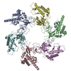
|
|---|---|
| 1 |
|
-Components
| #1: Protein | Mass: 93817.703 Da / Num. of mol.: 3 / Mutation: E519Q Source method: isolated from a genetically manipulated source Source: (gene. exp.) Homo sapiens (human) / Gene: MCM8, C20orf154 / Production host:  Trichoplusia ni (cabbage looper) / References: UniProt: Q9UJA3, DNA helicase Trichoplusia ni (cabbage looper) / References: UniProt: Q9UJA3, DNA helicase#2: Protein | Mass: 127485.594 Da / Num. of mol.: 3 / Mutation: E415Q Source method: isolated from a genetically manipulated source Source: (gene. exp.) Homo sapiens (human) / Gene: MCM9, C6orf61, MCMDC1 / Production host: Trichoplusia ni (cabbage looper) / References: UniProt: Q9NXL9, DNA helicase |
|---|
-Experimental details
-Experiment
| Experiment | Method: ELECTRON MICROSCOPY |
|---|---|
| EM experiment | Aggregation state: PARTICLE / 3D reconstruction method: single particle reconstruction |
- Sample preparation
| Component | Name: Structure of Walker B mutated MCM8/9 heterohexamer complex with ADP, N-terminal domains locally refined Type: COMPLEX / Entity ID: all / Source: RECOMBINANT |
|---|---|
| Molecular weight | Value: 0.500 MDa / Experimental value: NO |
| Source (natural) | Organism: Homo sapiens (human) |
| Source (recombinant) | Organism: Trichoplusia ni (cabbage looper) |
| Buffer solution | pH: 8 |
| Specimen | Conc.: 0.5 mg/ml / Embedding applied: NO / Shadowing applied: NO / Staining applied: NO / Vitrification applied: YES |
| Vitrification | Instrument: FEI VITROBOT MARK I / Cryogen name: ETHANE / Humidity: 100 % / Chamber temperature: 298 K |
- Electron microscopy imaging
| Experimental equipment |  Model: Titan Krios / Image courtesy: FEI Company |
|---|---|
| Microscopy | Model: FEI TITAN KRIOS |
| Electron gun | Electron source:  FIELD EMISSION GUN / Accelerating voltage: 300 kV / Illumination mode: FLOOD BEAM FIELD EMISSION GUN / Accelerating voltage: 300 kV / Illumination mode: FLOOD BEAM |
| Electron lens | Mode: BRIGHT FIELD / Nominal defocus max: 3000 nm / Nominal defocus min: 700 nm |
| Image recording | Electron dose: 49 e/Å2 / Film or detector model: GATAN K2 SUMMIT (4k x 4k) |
- Processing
| Software | Name: PHENIX / Version: 1.20.1_4487: / Classification: refinement | ||||||||||||||||||||||||
|---|---|---|---|---|---|---|---|---|---|---|---|---|---|---|---|---|---|---|---|---|---|---|---|---|---|
| CTF correction | Type: NONE | ||||||||||||||||||||||||
| 3D reconstruction | Resolution: 4.06 Å / Resolution method: FSC 0.143 CUT-OFF / Num. of particles: 227818 / Symmetry type: POINT | ||||||||||||||||||||||||
| Refine LS restraints |
|
