Movie
Movie Controller


+ Open data

- Basic information
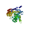
| Entry | Database: PDB / ID: 8r7h | |||||||||
|---|---|---|---|---|---|---|---|---|---|---|
| Title | Cryo-EM structure of Human SHMT1 | |||||||||
 Components Components | (Serine hydroxymethyltransferase, cytosolic) x 2 | |||||||||
 Keywords Keywords | TRANSFERASE / Riboregulation / Serine / Glycine Metabolism / 1 carbon metablism / Moonlighting protein / RNA BINDING PROTEIN | |||||||||
| Function / homology |  Function and homology information Function and homology informationphenylserine aldolase activity / threonine aldolase activity / cellular response to tetrahydrofolate / Carnitine synthesis / intracellular organelle lumen / hydroxytrimethyllysine aldolase activity / carnitine biosynthetic process / purine nucleobase biosynthetic process / serine binding / L-allo-threonine aldolase activity ...phenylserine aldolase activity / threonine aldolase activity / cellular response to tetrahydrofolate / Carnitine synthesis / intracellular organelle lumen / hydroxytrimethyllysine aldolase activity / carnitine biosynthetic process / purine nucleobase biosynthetic process / serine binding / L-allo-threonine aldolase activity / glycine metabolic process / L-serine catabolic process / L-serine metabolic process / aldehyde-lyase activity / glycine hydroxymethyltransferase / glycine hydroxymethyltransferase activity / : / lamin binding / Metabolism of folate and pterines / tetrahydrofolate metabolic process / tetrahydrofolate interconversion / dTMP biosynthetic process / small molecule binding / folic acid metabolic process / 'de novo' pyrimidine nucleobase biosynthetic process / one-carbon metabolic process / mRNA regulatory element binding translation repressor activity / replication fork / mRNA 5'-UTR binding / pyridoxal phosphate binding / protein homotetramerization / DNA replication / negative regulation of translation / protein homodimerization activity / mitochondrion / extracellular exosome / identical protein binding / nucleus / cytosol / cytoplasm Similarity search - Function | |||||||||
| Biological species |  Homo sapiens (human) Homo sapiens (human) | |||||||||
| Method | ELECTRON MICROSCOPY / single particle reconstruction / cryo EM / Resolution: 3.29 Å | |||||||||
 Authors Authors | Spizzichino, S. / Marabelli, C. / Bharadwaj, A. / Jakobi, A.J. / Chaves-Sanjuan, A. / Giardina, G. / Bolognesi, M. / Cutruzzola, F. | |||||||||
| Funding support |  Italy, 2items Italy, 2items
| |||||||||
 Citation Citation | Journal: Mol Cell / Year: 2024 Title: Structure-based mechanism of riboregulation of the metabolic enzyme SHMT1. Authors: Sharon Spizzichino / Federica Di Fonzo / Chiara Marabelli / Angela Tramonti / Antonio Chaves-Sanjuan / Alessia Parroni / Giovanna Boumis / Francesca Romana Liberati / Alessio Paone / Linda ...Authors: Sharon Spizzichino / Federica Di Fonzo / Chiara Marabelli / Angela Tramonti / Antonio Chaves-Sanjuan / Alessia Parroni / Giovanna Boumis / Francesca Romana Liberati / Alessio Paone / Linda Celeste Montemiglio / Matteo Ardini / Arjen J Jakobi / Alok Bharadwaj / Paolo Swuec / Gian Gaetano Tartaglia / Alessandro Paiardini / Roberto Contestabile / Antonello Mai / Dante Rotili / Francesco Fiorentino / Alberto Macone / Alessandra Giorgi / Giancarlo Tria / Serena Rinaldo / Martino Bolognesi / Giorgio Giardina / Francesca Cutruzzolà /  Abstract: RNA can directly control protein activity in a process called riboregulation; only a few mechanisms of riboregulation have been described in detail, none of which have been characterized on ...RNA can directly control protein activity in a process called riboregulation; only a few mechanisms of riboregulation have been described in detail, none of which have been characterized on structural grounds. Here, we present a comprehensive structural, functional, and phylogenetic analysis of riboregulation of cytosolic serine hydroxymethyltransferase (SHMT1), the enzyme interconverting serine and glycine in one-carbon metabolism. We have determined the cryoelectron microscopy (cryo-EM) structure of human SHMT1 in its free- and RNA-bound states, and we show that the RNA modulator competes with polyglutamylated folates and acts as an allosteric switch, selectively altering the enzyme's reactivity vs. serine. In addition, we identify the tetrameric assembly and a flap structural motif as key structural elements necessary for binding of RNA to eukaryotic SHMT1. The results presented here suggest that riboregulation may have played a role in evolution of eukaryotic SHMT1 and in compartmentalization of one-carbon metabolism. Our findings provide insights for RNA-based therapeutic strategies targeting this cancer-linked metabolic pathway. | |||||||||
| History |
|
- Structure visualization
| Structure viewer | Molecule: MolmilJmol/JSmol |
|---|
- Downloads & links
-Download
| PDBx/mmCIF format | 8r7h.cif.gz | 317.4 KB | Display | PDBx/mmCIF format |
|---|---|---|---|---|
| PDB format | pdb8r7h.ent.gz | 257.5 KB | Display | PDB format |
| PDBx/mmJSON format | 8r7h.json.gz | Tree view | PDBx/mmJSON format | |
| Others |  Other downloads Other downloads |
-Validation report
| Arichive directory | https://data.pdbj.org/pub/pdb/validation_reports/r7/8r7hftp://data.pdbj.org/pub/pdb/validation_reports/r7/8r7h | HTTPS FTP |
|---|
-Related structure data
| Related structure data |  18973MC  8a11C M: map data used to model this data C: citing same article ( |
|---|---|
| Similar structure data |

-Links
 PDBj
PDBj
- Assembly
| Deposited unit | 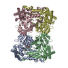
|
|---|---|
| 1 |
|
-Components
| #1: Protein | Mass: 53662.914 Da / Num. of mol.: 2 Source method: isolated from a genetically manipulated source Source: (gene. exp.) Homo sapiens (human) / Gene: SHMT1 / Production host:  References: UniProt: P34896, glycine hydroxymethyltransferase #2: Protein | Mass: 53434.797 Da / Num. of mol.: 2 Source method: isolated from a genetically manipulated source Source: (gene. exp.) Homo sapiens (human) / Gene: SHMT1 / Production host: Has ligand of interest | N | |
|---|
-Experimental details
-Experiment
| Experiment | Method: ELECTRON MICROSCOPY |
|---|---|
| EM experiment | Aggregation state: PARTICLE / 3D reconstruction method: single particle reconstruction |
- Sample preparation
| Component | Name: human SHMT1 / Type: COMPLEX / Details: SHMT1= homotetramer of 53kDa subunits / Entity ID: all / Source: RECOMBINANT | |||||||||||||||
|---|---|---|---|---|---|---|---|---|---|---|---|---|---|---|---|---|
| Molecular weight | Value: 0.21 MDa / Experimental value: NO | |||||||||||||||
| Source (natural) | Organism: Homo sapiens (human) | |||||||||||||||
| Source (recombinant) | Organism: | |||||||||||||||
| Buffer solution | pH: 7.2 | |||||||||||||||
| Buffer component |
| |||||||||||||||
| Specimen | Conc.: 0.3 mg/ml / Embedding applied: NO / Shadowing applied: NO / Staining applied: NO / Vitrification applied: YES | |||||||||||||||
| Vitrification | Instrument: FEI VITROBOT MARK IV / Cryogen name: ETHANE / Humidity: 100 % / Chamber temperature: 277 K / Details: blotted for 4 seconds before plunging |
- Electron microscopy imaging
| Experimental equipment |  Model: Talos Arctica / Image courtesy: FEI Company |
|---|---|
| Microscopy | Model: FEI TALOS ARCTICA |
| Electron gun | Electron source:  FIELD EMISSION GUN / Accelerating voltage: 200 kV / Illumination mode: FLOOD BEAM FIELD EMISSION GUN / Accelerating voltage: 200 kV / Illumination mode: FLOOD BEAM |
| Electron lens | Mode: BRIGHT FIELD / Nominal magnification: 120000 X / Nominal defocus max: 2500 nm / Nominal defocus min: 500 nm / Cs: 2.7 mm / Alignment procedure: BASIC |
| Specimen holder | Cryogen: NITROGEN / Specimen holder model: OTHER |
| Image recording | Electron dose: 40 e/Å2 / Detector mode: COUNTING / Film or detector model: FEI FALCON III (4k x 4k) / Num. of grids imaged: 1 / Num. of real images: 5450 |
- Processing
| EM software |
| ||||||||||||||||||||||||||||||||||||||||||||
|---|---|---|---|---|---|---|---|---|---|---|---|---|---|---|---|---|---|---|---|---|---|---|---|---|---|---|---|---|---|---|---|---|---|---|---|---|---|---|---|---|---|---|---|---|---|
| CTF correction | Type: PHASE FLIPPING AND AMPLITUDE CORRECTION | ||||||||||||||||||||||||||||||||||||||||||||
| Particle selection | Num. of particles selected: 4900000 | ||||||||||||||||||||||||||||||||||||||||||||
| 3D reconstruction | Resolution: 3.29 Å / Resolution method: FSC 0.143 CUT-OFF / Num. of particles: 94430 / Num. of class averages: 3 / Symmetry type: POINT | ||||||||||||||||||||||||||||||||||||||||||||
| Atomic model building | B value: 132 / Protocol: FLEXIBLE FIT / Space: REAL | ||||||||||||||||||||||||||||||||||||||||||||
| Atomic model building | PDB-ID: 1BJ4 Pdb chain-ID: A / Accession code: 1BJ4 / Source name: PDB / Type: experimental model | ||||||||||||||||||||||||||||||||||||||||||||
| Refine LS restraints |
|