Movie
Movie Controller

[English] 日本語
 Yorodumi
- PDB-8q6j: Atomic structure and conformational variability of the HER2-Trast... -
+ Open data

- Basic information
| Entry | Database: PDB / ID: 8q6j | |||||||||
|---|---|---|---|---|---|---|---|---|---|---|
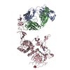
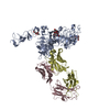
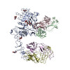
| Title | Atomic structure and conformational variability of the HER2-Trastuzumab-Pertuzumab complex | |||||||||
 Components Components |
| |||||||||
 Keywords Keywords | STRUCTURAL PROTEIN / ErbB-2 / Pertuzumab / Trastuzumab / Ternary complex / Protein / Flexibility / Continuous conformation / Cryo-EM / Single particle analysis | |||||||||
| Function / homology |  Function and homology information Function and homology informationnegative regulation of immature T cell proliferation in thymus / ERBB3:ERBB2 complex / ERBB2-ERBB4 signaling pathway / immature T cell proliferation in thymus / GRB7 events in ERBB2 signaling / RNA polymerase I core binding / semaphorin receptor complex / Developmental Lineage of Mammary Stem Cells / ErbB-3 class receptor binding / Sema4D induced cell migration and growth-cone collapse ...negative regulation of immature T cell proliferation in thymus / ERBB3:ERBB2 complex / ERBB2-ERBB4 signaling pathway / immature T cell proliferation in thymus / GRB7 events in ERBB2 signaling / RNA polymerase I core binding / semaphorin receptor complex / Developmental Lineage of Mammary Stem Cells / ErbB-3 class receptor binding / Sema4D induced cell migration and growth-cone collapse / motor neuron axon guidance / regulation of microtubule-based process / PLCG1 events in ERBB2 signaling / enzyme-linked receptor protein signaling pathway / ERBB2 Activates PTK6 Signaling / ERBB2-EGFR signaling pathway / ERBB2-ERBB3 signaling pathway / neurotransmitter receptor localization to postsynaptic specialization membrane / Drug-mediated inhibition of ERBB2 signaling / Resistance of ERBB2 KD mutants to trastuzumab / Resistance of ERBB2 KD mutants to sapitinib / Resistance of ERBB2 KD mutants to tesevatinib / Resistance of ERBB2 KD mutants to neratinib / Resistance of ERBB2 KD mutants to osimertinib / Resistance of ERBB2 KD mutants to afatinib / Resistance of ERBB2 KD mutants to AEE788 / Resistance of ERBB2 KD mutants to lapatinib / Drug resistance in ERBB2 TMD/JMD mutants / positive regulation of MAP kinase activity / positive regulation of Rho protein signal transduction / neuromuscular junction development / positive regulation of transcription by RNA polymerase I / ERBB2 Regulates Cell Motility / Developmental Lineage of Mammary Gland Myoepithelial Cells / oligodendrocyte differentiation / semaphorin-plexin signaling pathway / PI3K events in ERBB2 signaling / regulation of angiogenesis / positive regulation of protein targeting to membrane / regulation of ERK1 and ERK2 cascade / Schwann cell development / coreceptor activity / Signaling by ERBB2 / peptidyl-tyrosine phosphorylation / TFAP2 (AP-2) family regulates transcription of growth factors and their receptors / myelination / transmembrane receptor protein tyrosine kinase activity / GRB2 events in ERBB2 signaling / positive regulation of cell adhesion / SHC1 events in ERBB2 signaling / cell surface receptor protein tyrosine kinase signaling pathway / basal plasma membrane / cellular response to epidermal growth factor stimulus / Constitutive Signaling by Overexpressed ERBB2 / Downregulation of ERBB2:ERBB3 signaling / positive regulation of epithelial cell proliferation / positive regulation of translation / neuromuscular junction / wound healing / phosphatidylinositol 3-kinase/protein kinase B signal transduction / Signaling by ERBB2 TMD/JMD mutants / receptor protein-tyrosine kinase / Signaling by ERBB2 ECD mutants / Signaling by ERBB2 KD Mutants / receptor tyrosine kinase binding / cellular response to growth factor stimulus / Downregulation of ERBB2 signaling / ruffle membrane / epidermal growth factor receptor signaling pathway / neuron differentiation / Constitutive Signaling by Aberrant PI3K in Cancer / transmembrane signaling receptor activity / PIP3 activates AKT signaling / myelin sheath / heart development / PI5P, PP2A and IER3 Regulate PI3K/AKT Signaling / RAF/MAP kinase cascade / positive regulation of cell growth / protein tyrosine kinase activity / presynaptic membrane / basolateral plasma membrane / protein phosphorylation / early endosome / positive regulation of MAPK cascade / cell surface receptor signaling pathway / signaling receptor complex / cell population proliferation / endosome membrane / apical plasma membrane / intracellular signal transduction / protein heterodimerization activity / signaling receptor binding / negative regulation of apoptotic process / perinuclear region of cytoplasm / signal transduction / nucleoplasm / ATP binding / membrane / identical protein binding / nucleus Similarity search - Function | |||||||||
| Biological species |  Homo sapiens (human) Homo sapiens (human) | |||||||||
| Method | ELECTRON MICROSCOPY / single particle reconstruction / cryo EM / Resolution: 3.3 Å | |||||||||
 Authors Authors | Ruedas, R. / Vuillemot, R. / Tubiana, T. / Winter, J.M. / Pieri, L. / Arteni, A.A. / Samson, C. / Jonic, J. / Mathieu, M. / Bressanelli, S. | |||||||||
| Funding support |  France, 2items France, 2items
| |||||||||
 Citation Citation | Journal: J Struct Biol / Year: 2024 Title: Structure and conformational variability of the HER2-trastuzumab-pertuzumab complex. Authors: Rémi Ruedas / Rémi Vuillemot / Thibault Tubiana / Jean-Marie Winter / Laura Pieri / Ana-Andreea Arteni / Camille Samson / Slavica Jonic / Magali Mathieu / Stéphane Bressanelli / Abstract: Single particle analysis from cryogenic transmission electron microscopy (cryo-EM) is particularly attractive for complexes for which structure prediction remains intractable, such as antibody- ...Single particle analysis from cryogenic transmission electron microscopy (cryo-EM) is particularly attractive for complexes for which structure prediction remains intractable, such as antibody-antigen complexes. Here we obtain the detailed structure of a particularly difficult complex between human epidermal growth factor receptor 2 (HER2) and the antigen-binding fragments from two distinct therapeutic antibodies binding to distant parts of the flexible HER2, pertuzumab and trastuzumab (HTP). We highlight the strengths and limitations of current data processing software in dealing with various kinds of heterogeneities, particularly continuous conformational heterogeneity, and in describing the motions that can be extracted from our dataset. Our HTP structure provides a more detailed view than the one previously available for this ternary complex. This allowed us to pinpoint a previously overlooked loop in domain IV that may be involved both in binding of trastuzumab and in HER2 dimerization. This finding may contribute to explain the synergistic anticancer effect of the two antibodies. We further propose that the flexibility of the HTP complex, beyond the difficulties it causes for cryo-EM analysis, actually reflects regulation of HER2 signaling and its inhibition by therapeutic antibodies. Notably we obtain our best data with ultra-thin continuous carbon grids, showing that with current cameras their use to alleviate particle misdistribution is compatible with a protein complex of only 162 kDa. Perhaps most importantly, we provide here a dataset for such a smallish protein complex for further development of software accounting for continuous conformational heterogeneity in cryo-EM images. | |||||||||
| History |
|
- Structure visualization
| Structure viewer | Molecule: MolmilJmol/JSmol |
|---|
- Downloads & links
-Download
| PDBx/mmCIF format | 8q6j.cif.gz | 299.2 KB | Display | PDBx/mmCIF format |
|---|---|---|---|---|
| PDB format | pdb8q6j.ent.gz | Display | PDB format | |
| PDBx/mmJSON format | 8q6j.json.gz | Tree view | PDBx/mmJSON format | |
| Others |  Other downloads Other downloads |
-Validation report
| Arichive directory | https://data.pdbj.org/pub/pdb/validation_reports/q6/8q6jftp://data.pdbj.org/pub/pdb/validation_reports/q6/8q6j | HTTPS FTP |
|---|
-Related structure data
| Related structure data | 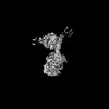 18188MC 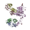 8pwhC M: map data used to model this data C: citing same article ( |
|---|---|
| Similar structure data |

-Links
 PDBj
PDBj


- Assembly
| Deposited unit | 
|
|---|---|
| 1 |
|
-Components
-Protein , 1 types, 1 molecules E
| #5: Protein | Mass: 68794.086 Da / Num. of mol.: 1 Source method: isolated from a genetically manipulated source Source: (gene. exp.) Homo sapiens (human) / Gene: ERBB2 / Cell line (production host): HEK293 / Production host: Homo sapiens (human) / References: UniProt: P04626 |
|---|
-Antibody , 4 types, 4 molecules ABCD
| #1: Antibody | Mass: 23466.031 Da / Num. of mol.: 1 Source method: isolated from a genetically manipulated source Source: (gene. exp.) Homo sapiens (human) / Production host:   Cricetulus griseus (Chinese hamster) Cricetulus griseus (Chinese hamster) |
|---|---|
| #2: Antibody | Mass: 23425.180 Da / Num. of mol.: 1 Source method: isolated from a genetically manipulated source Source: (gene. exp.) Homo sapiens (human) / Production host: Cricetulus griseus (Chinese hamster) |
| #3: Antibody | Mass: 23548.152 Da / Num. of mol.: 1 Source method: isolated from a genetically manipulated source Source: (gene. exp.) Homo sapiens (human) / Production host: Cricetulus griseus (Chinese hamster) |
| #4: Antibody | Mass: 23674.486 Da / Num. of mol.: 1 Source method: isolated from a genetically manipulated source Source: (gene. exp.) Homo sapiens (human) / Cell line (production host): CHO / Production host: Cricetulus griseus (Chinese hamster) |
-Sugars , 2 types, 5 molecules 
| #6: Polysaccharide | Source method: isolated from a genetically manipulated source #7: Sugar |  Type: D-saccharide, beta linking / Mass: 221.208 Da / Num. of mol.: 3 Type: D-saccharide, beta linking / Mass: 221.208 Da / Num. of mol.: 3Source method: isolated from a genetically manipulated source Formula: C8H15NO6 |
|---|
-Details
| Has ligand of interest | N |
|---|---|
| Has protein modification | Y |
-Experimental details
-Experiment
| Experiment | Method: ELECTRON MICROSCOPY |
|---|---|
| EM experiment | Aggregation state: PARTICLE / 3D reconstruction method: single particle reconstruction |
- Sample preparation
| Component | Name: Ternary complex of her2-trastuzumab-pertuzumab Fabs / Type: COMPLEX Details: Fab fragment from trastuzumab and pertuzumab linked to the extracellular domain of erbb2 (her2) Entity ID: #1-#4 / Source: RECOMBINANT | |||||||||||||||
|---|---|---|---|---|---|---|---|---|---|---|---|---|---|---|---|---|
| Molecular weight | Value: 0.171364 MDa / Experimental value: YES | |||||||||||||||
| Source (natural) | Organism: Homo sapiens (human) | |||||||||||||||
| Source (recombinant) | Organism: Homo sapiens (human) / Strain: HEK293 / Cell: human | |||||||||||||||
| Buffer solution | pH: 7.5 | |||||||||||||||
| Buffer component |
| |||||||||||||||
| Specimen | Conc.: 0.12 mg/ml / Embedding applied: NO / Shadowing applied: NO / Staining applied: NO / Vitrification applied: YES | |||||||||||||||
| Vitrification | Instrument: FEI VITROBOT MARK IV / Cryogen name: ETHANE / Humidity: 100 % / Chamber temperature: 288.15 K |
- Electron microscopy imaging
| Microscopy | Model: TFS GLACIOS |
|---|---|
| Electron gun | Electron source:  FIELD EMISSION GUN / Accelerating voltage: 200 kV / Illumination mode: FLOOD BEAM FIELD EMISSION GUN / Accelerating voltage: 200 kV / Illumination mode: FLOOD BEAM |
| Electron lens | Mode: BRIGHT FIELD / Nominal magnification: 240000 X / Nominal defocus max: 2600 nm / Nominal defocus min: 800 nm / Cs: 2.1 mm / Alignment procedure: BASIC |
| Specimen holder | Cryogen: HELIUM / Specimen holder model: FEI TITAN KRIOS AUTOGRID HOLDER |
| Image recording | Average exposure time: 4.71 sec. / Electron dose: 60 e/Å2 / Film or detector model: FEI FALCON IV (4k x 4k) / Num. of grids imaged: 1 / Num. of real images: 8928 |
| Image scans | Width: 4096 / Height: 4096 |
- Processing
| EM software |
| |||||||||||||||||||||||||||||||||||||||||||||||||||||||||||||||||
|---|---|---|---|---|---|---|---|---|---|---|---|---|---|---|---|---|---|---|---|---|---|---|---|---|---|---|---|---|---|---|---|---|---|---|---|---|---|---|---|---|---|---|---|---|---|---|---|---|---|---|---|---|---|---|---|---|---|---|---|---|---|---|---|---|---|---|
| CTF correction | Type: PHASE FLIPPING AND AMPLITUDE CORRECTION | |||||||||||||||||||||||||||||||||||||||||||||||||||||||||||||||||
| Particle selection | Num. of particles selected: 2954825 | |||||||||||||||||||||||||||||||||||||||||||||||||||||||||||||||||
| 3D reconstruction | Resolution: 3.3 Å / Resolution method: FSC 0.143 CUT-OFF / Num. of particles: 711008 / Algorithm: FOURIER SPACE / Symmetry type: POINT | |||||||||||||||||||||||||||||||||||||||||||||||||||||||||||||||||
| Atomic model building | Protocol: RIGID BODY FIT / Space: REAL | |||||||||||||||||||||||||||||||||||||||||||||||||||||||||||||||||
| Atomic model building | 3D fitting-ID: 1
| |||||||||||||||||||||||||||||||||||||||||||||||||||||||||||||||||
| Refine LS restraints |
|