Movie
Movie Controller

[English] 日本語
 Yorodumi
- PDB-8piu: 60-meric complex of dihydrolipoamide acetyltransferase (E2) of th... -
+ Open data

- Basic information
| Entry | Database: PDB / ID: 8piu | ||||||
|---|---|---|---|---|---|---|---|
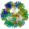
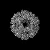
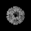
| Title | 60-meric complex of dihydrolipoamide acetyltransferase (E2) of the human pyruvate dehydrogenase complex | ||||||
 Components Components | Dihydrolipoyllysine-residue acetyltransferase component of pyruvate dehydrogenase complex, mitochondrial | ||||||
 Keywords Keywords | TRANSFERASE / pyruvate dehydrogenase complex / PDHc / E2 / cryo-EM | ||||||
| Function / homology |  Function and homology information Function and homology informationPDH complex synthesizes acetyl-CoA from PYR / dihydrolipoyllysine-residue acetyltransferase / dihydrolipoyllysine-residue acetyltransferase activity / Regulation of pyruvate dehydrogenase (PDH) complex / Protein lipoylation / pyruvate decarboxylation to acetyl-CoA / pyruvate catabolic process / pyruvate dehydrogenase complex / Signaling by Retinoic Acid / sperm head-tail coupling apparatus ...PDH complex synthesizes acetyl-CoA from PYR / dihydrolipoyllysine-residue acetyltransferase / dihydrolipoyllysine-residue acetyltransferase activity / Regulation of pyruvate dehydrogenase (PDH) complex / Protein lipoylation / pyruvate decarboxylation to acetyl-CoA / pyruvate catabolic process / pyruvate dehydrogenase complex / Signaling by Retinoic Acid / sperm head-tail coupling apparatus / tricarboxylic acid cycle / glucose metabolic process / mitochondrial matrix / mitochondrion / identical protein binding Similarity search - Function | ||||||
| Biological species |  Homo sapiens (human) Homo sapiens (human) | ||||||
| Method | ELECTRON MICROSCOPY / single particle reconstruction / cryo EM / Resolution: 2.9 Å | ||||||
 Authors Authors | Zdanowicz, R. / Afanasyev, P. / Boehringer, D. / Glockshuber, R. | ||||||
| Funding support |  Switzerland, 1items Switzerland, 1items
| ||||||
 Citation Citation | Journal: Sci Adv / Year: 2024 Title: Stoichiometry and architecture of the human pyruvate dehydrogenase complex. Authors: Rafal Zdanowicz / Pavel Afanasyev / Adam Pruška / Julian A Harrison / Christoph Giese / Daniel Boehringer / Alexander Leitner / Renato Zenobi / Rudi Glockshuber / Abstract: The pyruvate dehydrogenase complex (PDHc) is a key megaenzyme linking glycolysis with the citric acid cycle. In mammalian PDHc, dihydrolipoamide acetyltransferase (E2) and the dihydrolipoamide ...The pyruvate dehydrogenase complex (PDHc) is a key megaenzyme linking glycolysis with the citric acid cycle. In mammalian PDHc, dihydrolipoamide acetyltransferase (E2) and the dihydrolipoamide dehydrogenase-binding protein (E3BP) form a 60-subunit core that associates with the peripheral subunits pyruvate dehydrogenase (E1) and dihydrolipoamide dehydrogenase (E3). The structure and stoichiometry of the fully assembled, mammalian PDHc or its core remained elusive. Here, we demonstrate that the human PDHc core is formed by 48 E2 copies that bind 48 E1 heterotetramers and 12 E3BP copies that bind 12 E3 homodimers. Cryo-electron microscopy, together with native and cross-linking mass spectrometry, confirmed a core model in which 8 E2 homotrimers and 12 E2-E2-E3BP heterotrimers assemble into a pseudoicosahedral particle such that the 12 E3BP molecules form six E3BP-E3BP intertrimer interfaces distributed tetrahedrally within the 60-subunit core. The even distribution of E3 subunits in the peripheral shell of PDHc guarantees maximum enzymatic activity of the megaenzyme. | ||||||
| History |
|
- Structure visualization
| Structure viewer | Molecule: MolmilJmol/JSmol |
|---|
- Downloads & links
-Download
| PDBx/mmCIF format | 8piu.cif.gz | 2.6 MB | Display | PDBx/mmCIF format |
|---|---|---|---|---|
| PDB format | pdb8piu.ent.gz | Display | PDB format | |
| PDBx/mmJSON format | 8piu.json.gz | Tree view | PDBx/mmJSON format | |
| Others |  Other downloads Other downloads |
-Validation report
| Arichive directory | https://data.pdbj.org/pub/pdb/validation_reports/pi/8piuftp://data.pdbj.org/pub/pdb/validation_reports/pi/8piu | HTTPS FTP |
|---|
-Related structure data
| Related structure data | 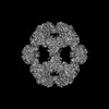 17691MC M: map data used to model this data C: citing same article ( |
|---|---|
| Similar structure data |

-Links
 PDBj
PDBj


- Assembly
| Deposited unit | 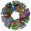
|
|---|---|
| 1 |
|
-Components
| #1: Protein | Mass: 59811.832 Da / Num. of mol.: 60 Source method: isolated from a genetically manipulated source Source: (gene. exp.) Homo sapiens (human) / Gene: DLAT, DLTA / Production host:  References: UniProt: P10515, dihydrolipoyllysine-residue acetyltransferase |
|---|
-Experimental details
-Experiment
| Experiment | Method: ELECTRON MICROSCOPY |
|---|---|
| EM experiment | Aggregation state: PARTICLE / 3D reconstruction method: single particle reconstruction |
- Sample preparation
| Component | Name: 60-meric complex of dihydrolipoamide acetyltransferase (E2) of the human pyruvate dehydrogenase complex Type: COMPLEX / Entity ID: all / Source: RECOMBINANT |
|---|---|
| Molecular weight | Value: 3.6 MDa / Experimental value: NO |
| Source (natural) | Organism: Homo sapiens (human) |
| Source (recombinant) | Organism: |
| Buffer solution | pH: 7.4 |
| Specimen | Embedding applied: NO / Shadowing applied: NO / Staining applied: NO / Vitrification applied: YES |
| Specimen support | Grid material: COPPER / Grid type: Quantifoil R2/2 |
| Vitrification | Instrument: FEI VITROBOT MARK IV / Cryogen name: ETHANE / Humidity: 100 % / Chamber temperature: 277 K |
- Electron microscopy imaging
| Experimental equipment |  Model: Titan Krios / Image courtesy: FEI Company |
|---|---|
| Microscopy | Model: FEI TITAN KRIOS |
| Electron gun | Electron source:  FIELD EMISSION GUN / Accelerating voltage: 300 kV / Illumination mode: FLOOD BEAM FIELD EMISSION GUN / Accelerating voltage: 300 kV / Illumination mode: FLOOD BEAM |
| Electron lens | Mode: BRIGHT FIELD / Nominal defocus max: 2800 nm / Nominal defocus min: 1600 nm |
| Image recording | Electron dose: 80 e/Å2 / Detector mode: COUNTING / Film or detector model: GATAN K2 SUMMIT (4k x 4k) |
- Processing
| EM software |
| ||||||||||||||||||||||||||||||||||||||||
|---|---|---|---|---|---|---|---|---|---|---|---|---|---|---|---|---|---|---|---|---|---|---|---|---|---|---|---|---|---|---|---|---|---|---|---|---|---|---|---|---|---|
| CTF correction | Type: PHASE FLIPPING AND AMPLITUDE CORRECTION | ||||||||||||||||||||||||||||||||||||||||
| Symmetry | Point symmetry: I (icosahedral) | ||||||||||||||||||||||||||||||||||||||||
| 3D reconstruction | Resolution: 2.9 Å / Resolution method: FSC 0.143 CUT-OFF / Num. of particles: 83959 / Symmetry type: POINT | ||||||||||||||||||||||||||||||||||||||||
| Atomic model building | PDB-ID: 6CT0 Accession code: 6CT0 / Source name: PDB / Type: experimental model |