[English] 日本語
 Yorodumi
- PDB-8p5d: Spraguea lophii ribosome in the closed conformation by cryo sub t... -
+ Open data
- Basic information
| Entry | Database: PDB / ID: 8p5d | ||||||
|---|---|---|---|---|---|---|---|
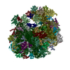
| Title | Spraguea lophii ribosome in the closed conformation by cryo sub tomogram averaging | ||||||
 Components Components |
| ||||||
 Keywords Keywords | RIBOSOME / Microsporidia | ||||||
| Function / homology |  Function and homology information Function and homology information90S preribosome / translation regulator activity / maturation of LSU-rRNA / maturation of LSU-rRNA from tricistronic rRNA transcript (SSU-rRNA, 5.8S rRNA, LSU-rRNA) / rescue of stalled ribosome / ribosomal large subunit biogenesis / maturation of SSU-rRNA from tricistronic rRNA transcript (SSU-rRNA, 5.8S rRNA, LSU-rRNA) / protein kinase C binding / modification-dependent protein catabolic process / protein tag activity ...90S preribosome / translation regulator activity / maturation of LSU-rRNA / maturation of LSU-rRNA from tricistronic rRNA transcript (SSU-rRNA, 5.8S rRNA, LSU-rRNA) / rescue of stalled ribosome / ribosomal large subunit biogenesis / maturation of SSU-rRNA from tricistronic rRNA transcript (SSU-rRNA, 5.8S rRNA, LSU-rRNA) / protein kinase C binding / modification-dependent protein catabolic process / protein tag activity / ribosomal small subunit biogenesis / small ribosomal subunit rRNA binding / ribosome binding / ribosomal small subunit assembly / small ribosomal subunit / 5S rRNA binding / large ribosomal subunit rRNA binding / cytosolic small ribosomal subunit / ribosomal large subunit assembly / cytoplasmic translation / cytosolic large ribosomal subunit / negative regulation of translation / rRNA binding / ribosome / protein ubiquitination / structural constituent of ribosome / ribonucleoprotein complex / positive regulation of protein phosphorylation / translation / mRNA binding / ubiquitin protein ligase binding / nucleolus / RNA binding / zinc ion binding / membrane / nucleus / cytosol / cytoplasm Similarity search - Function | ||||||
| Biological species |  Spraguea lophii 42_110 (fungus) Spraguea lophii 42_110 (fungus) | ||||||
| Method | ELECTRON MICROSCOPY / subtomogram averaging / cryo EM / Resolution: 10.8 Å | ||||||
 Authors Authors | Gil Diez, P. / McLaren, M. / Isupov, M.N. / Daum, B. / Conners, R. / Williams, B. | ||||||
| Funding support |  United Kingdom, 1items United Kingdom, 1items
| ||||||
 Citation Citation |  Journal: Nat Microbiol / Year: 2023 Journal: Nat Microbiol / Year: 2023Title: CryoEM reveals that ribosomes in microsporidian spores are locked in a dimeric hibernating state. Authors: Mathew McLaren / Rebecca Conners / Michail N Isupov / Patricia Gil-Díez / Lavinia Gambelli / Vicki A M Gold / Andreas Walter / Sean R Connell / Bryony Williams / Bertram Daum /    Abstract: Translational control is an essential process for the cell to adapt to varying physiological or environmental conditions. To survive adverse conditions such as low nutrient levels, translation can be ...Translational control is an essential process for the cell to adapt to varying physiological or environmental conditions. To survive adverse conditions such as low nutrient levels, translation can be shut down almost entirely by inhibiting ribosomal function. Here we investigated eukaryotic hibernating ribosomes from the microsporidian parasite Spraguea lophii in situ by a combination of electron cryo-tomography and single-particle electron cryo-microscopy. We show that microsporidian spores contain hibernating ribosomes that are locked in a dimeric (100S) state, which is formed by a unique dimerization mechanism involving the beak region. The ribosomes within the dimer are fully assembled, suggesting that they are ready to be activated once the host cell is invaded. This study provides structural evidence for dimerization acting as a mechanism for ribosomal hibernation in microsporidia, and therefore demonstrates that eukaryotes utilize this mechanism in translational control. | ||||||
| History |
|
- Structure visualization
| Structure viewer | Molecule:  Molmil Molmil Jmol/JSmol Jmol/JSmol |
|---|
- Downloads & links
- Download
| PDBx/mmCIF format |  8p5d.cif.gz 8p5d.cif.gz | 3.7 MB | Display |  PDBx/mmCIF format PDBx/mmCIF format |
|---|---|---|---|---|
| PDB format |  pdb8p5d.ent.gz pdb8p5d.ent.gz | Display |  PDB format PDB format | |
| PDBx/mmJSON format |  8p5d.json.gz 8p5d.json.gz | Tree view |  PDBx/mmJSON format PDBx/mmJSON format | |
| Others |  Other downloads Other downloads |
-Validation report
| Arichive directory |  https://data.pdbj.org/pub/pdb/validation_reports/p5/8p5d https://data.pdbj.org/pub/pdb/validation_reports/p5/8p5d ftp://data.pdbj.org/pub/pdb/validation_reports/p5/8p5d ftp://data.pdbj.org/pub/pdb/validation_reports/p5/8p5d | HTTPS FTP |
|---|
-Related structure data
| Related structure data |  17448MC  8p60C  16198  7qjh M: map data used to model this data C: citing same article ( |
|---|---|
| Similar structure data | Similarity search - Function & homology  F&H Search F&H Search |
- Links
Links
- Assembly
| Deposited unit | 
|
|---|---|
| 1 |
|
- Components
-RNA chain , 3 types, 3 molecules L50L70S60
| #1: RNA chain | Mass: 849039.812 Da / Num. of mol.: 1 / Source method: isolated from a natural source / Source: (natural)  Spraguea lophii 42_110 (fungus) Spraguea lophii 42_110 (fungus) |
|---|---|
| #2: RNA chain | Mass: 38356.801 Da / Num. of mol.: 1 / Source method: isolated from a natural source / Source: (natural)  Spraguea lophii 42_110 (fungus) Spraguea lophii 42_110 (fungus) |
| #42: RNA chain | Mass: 444812.531 Da / Num. of mol.: 1 / Source method: isolated from a natural source / Source: (natural)  Spraguea lophii 42_110 (fungus) Spraguea lophii 42_110 (fungus) |
+60S ribosomal protein ... , 27 types, 27 molecules LA0LB0LC0LCCLD0LDDLE0LEELF0LFFLG0LH0LIILJ0LL0LLLLOOLP0LPPLQ0LR0LS0LT0LU0LX0LY0LZ0
-Protein , 15 types, 15 molecules LAALHHLI0LJJLM0LMMMD1SB0SBBSCCSEESFFSGGSR0SX0
| #4: Protein | Mass: 16609.498 Da / Num. of mol.: 1 / Source method: isolated from a natural source / Source: (natural)  Spraguea lophii 42_110 (fungus) Spraguea lophii 42_110 (fungus) |
|---|---|
| #17: Protein | Mass: 14183.866 Da / Num. of mol.: 1 / Source method: isolated from a natural source / Source: (natural)  Spraguea lophii 42_110 (fungus) / References: UniProt: S7W7Y2 Spraguea lophii 42_110 (fungus) / References: UniProt: S7W7Y2 |
| #18: Protein | Mass: 25142.328 Da / Num. of mol.: 1 / Source method: isolated from a natural source / Source: (natural)  Spraguea lophii 42_110 (fungus) / References: UniProt: S7WBF8 Spraguea lophii 42_110 (fungus) / References: UniProt: S7WBF8 |
| #21: Protein | Mass: 10517.657 Da / Num. of mol.: 1 / Source method: isolated from a natural source / Source: (natural)  Spraguea lophii 42_110 (fungus) Spraguea lophii 42_110 (fungus) |
| #24: Protein | Mass: 13258.343 Da / Num. of mol.: 1 / Source method: isolated from a natural source / Source: (natural)  Spraguea lophii 42_110 (fungus) / References: UniProt: S7XVN9 Spraguea lophii 42_110 (fungus) / References: UniProt: S7XVN9 |
| #25: Protein | Mass: 14563.868 Da / Num. of mol.: 1 / Source method: isolated from a natural source / Source: (natural)  Spraguea lophii 42_110 (fungus) / References: UniProt: S7XSQ3 Spraguea lophii 42_110 (fungus) / References: UniProt: S7XSQ3 |
| #41: Protein | Mass: 17595.088 Da / Num. of mol.: 1 / Source method: isolated from a natural source / Source: (natural)  Spraguea lophii 42_110 (fungus) / References: UniProt: S7W5K5 Spraguea lophii 42_110 (fungus) / References: UniProt: S7W5K5 |
| #45: Protein | Mass: 25934.658 Da / Num. of mol.: 1 / Source method: isolated from a natural source / Source: (natural)  Spraguea lophii 42_110 (fungus) Spraguea lophii 42_110 (fungus) |
| #46: Protein | Mass: 9189.923 Da / Num. of mol.: 1 / Source method: isolated from a natural source / Source: (natural)  Spraguea lophii 42_110 (fungus) Spraguea lophii 42_110 (fungus) |
| #48: Protein | Mass: 7288.479 Da / Num. of mol.: 1 / Source method: isolated from a natural source / Source: (natural)  Spraguea lophii 42_110 (fungus) Spraguea lophii 42_110 (fungus) |
| #52: Protein | Mass: 6816.010 Da / Num. of mol.: 1 / Source method: isolated from a natural source / Source: (natural)  Spraguea lophii 42_110 (fungus) Spraguea lophii 42_110 (fungus) |
| #54: Protein | Mass: 16937.791 Da / Num. of mol.: 1 / Source method: isolated from a natural source / Source: (natural)  Spraguea lophii 42_110 (fungus) / References: UniProt: S7W9X5 Spraguea lophii 42_110 (fungus) / References: UniProt: S7W9X5 |
| #56: Protein | Mass: 36182.824 Da / Num. of mol.: 1 / Source method: isolated from a natural source / Source: (natural)  Spraguea lophii 42_110 (fungus) / References: UniProt: S7XVG3 Spraguea lophii 42_110 (fungus) / References: UniProt: S7XVG3 |
| #67: Protein | Mass: 14055.251 Da / Num. of mol.: 1 / Source method: isolated from a natural source / Source: (natural)  Spraguea lophii 42_110 (fungus) Spraguea lophii 42_110 (fungus) |
| #73: Protein | Mass: 15822.722 Da / Num. of mol.: 1 / Source method: isolated from a natural source / Source: (natural)  Spraguea lophii 42_110 (fungus) Spraguea lophii 42_110 (fungus) |
-Ribosomal protein ... , 7 types, 7 molecules LGGLN0LO0LV0LW0SP0SV0
| #15: Protein | Mass: 12020.348 Da / Num. of mol.: 1 / Source method: isolated from a natural source / Source: (natural)  Spraguea lophii 42_110 (fungus) / References: UniProt: S7W7C6 Spraguea lophii 42_110 (fungus) / References: UniProt: S7W7C6 |
|---|---|
| #26: Protein | Mass: 24200.426 Da / Num. of mol.: 1 / Source method: isolated from a natural source / Source: (natural)  Spraguea lophii 42_110 (fungus) / References: UniProt: S7W7A2 Spraguea lophii 42_110 (fungus) / References: UniProt: S7W7A2 |
| #27: Protein | Mass: 22885.139 Da / Num. of mol.: 1 / Source method: isolated from a natural source / Source: (natural)  Spraguea lophii 42_110 (fungus) / References: UniProt: S7XUD8 Spraguea lophii 42_110 (fungus) / References: UniProt: S7XUD8 |
| #36: Protein | Mass: 15239.987 Da / Num. of mol.: 1 / Source method: isolated from a natural source / Source: (natural)  Spraguea lophii 42_110 (fungus) / References: UniProt: S7XLC4 Spraguea lophii 42_110 (fungus) / References: UniProt: S7XLC4 |
| #37: Protein | Mass: 15342.161 Da / Num. of mol.: 1 / Source method: isolated from a natural source / Source: (natural)  Spraguea lophii 42_110 (fungus) / References: UniProt: S7XSY2 Spraguea lophii 42_110 (fungus) / References: UniProt: S7XSY2 |
| #65: Protein | Mass: 18514.629 Da / Num. of mol.: 1 / Source method: isolated from a natural source / Source: (natural)  Spraguea lophii 42_110 (fungus) / References: UniProt: S7XKY9 Spraguea lophii 42_110 (fungus) / References: UniProt: S7XKY9 |
| #71: Protein | Mass: 7768.887 Da / Num. of mol.: 1 / Source method: isolated from a natural source / Source: (natural)  Spraguea lophii 42_110 (fungus) / References: UniProt: S7WAC1 Spraguea lophii 42_110 (fungus) / References: UniProt: S7WAC1 |
+40S ribosomal protein ... , 23 types, 23 molecules SA0SAASC0SD0SDDSE0SF0SG0SH0SI0SJ0SK0SL0SM0SN0SO0SQ0SS0ST0SU0SW0SY0SZ0
-Non-polymers , 1 types, 9 molecules 
| #76: Chemical | ChemComp-ZN / |
|---|
-Details
| Has ligand of interest | Y |
|---|
-Experimental details
-Experiment
| Experiment | Method: ELECTRON MICROSCOPY |
|---|---|
| EM experiment | Aggregation state: PARTICLE / 3D reconstruction method: subtomogram averaging |
- Sample preparation
| Component | Name: Ribosome / Type: RIBOSOME / Entity ID: #1-#64, #66-#75, #65 / Source: NATURAL |
|---|---|
| Source (natural) | Organism:  Spraguea lophii 42_110 (fungus) Spraguea lophii 42_110 (fungus) |
| Buffer solution | pH: 7.5 |
| Specimen | Embedding applied: NO / Shadowing applied: NO / Staining applied: NO / Vitrification applied: YES |
| Specimen support | Details: 20 mA, Carbon coated grid / Grid material: COPPER / Grid mesh size: 300 divisions/in. / Grid type: Quantifoil |
| Vitrification | Instrument: FEI VITROBOT MARK IV / Cryogen name: ETHANE / Humidity: 100 % / Chamber temperature: 288.15 K / Details: blot force -1 and blot time 4 s |
- Electron microscopy imaging
| Experimental equipment |  Model: Titan Krios / Image courtesy: FEI Company | ||||||||||||||||||
|---|---|---|---|---|---|---|---|---|---|---|---|---|---|---|---|---|---|---|---|
| Microscopy | Model: FEI TITAN KRIOS | ||||||||||||||||||
| Electron gun | Electron source:  FIELD EMISSION GUN / Accelerating voltage: 300 kV / Illumination mode: FLOOD BEAM FIELD EMISSION GUN / Accelerating voltage: 300 kV / Illumination mode: FLOOD BEAM | ||||||||||||||||||
| Electron lens | Mode: BRIGHT FIELD / Nominal defocus max: 6000 nm / Nominal defocus min: 2500 nm / Cs: 2.7 mm | ||||||||||||||||||
| Specimen holder | Cryogen: NITROGEN / Specimen holder model: FEI TITAN KRIOS AUTOGRID HOLDER | ||||||||||||||||||
| Image recording |
|
- Processing
| EM software |
| ||||||||||||||||||||||||||||||||
|---|---|---|---|---|---|---|---|---|---|---|---|---|---|---|---|---|---|---|---|---|---|---|---|---|---|---|---|---|---|---|---|---|---|
| CTF correction | Type: PHASE FLIPPING AND AMPLITUDE CORRECTION | ||||||||||||||||||||||||||||||||
| 3D reconstruction | Resolution: 10.8 Å / Resolution method: FSC 0.143 CUT-OFF / Num. of particles: 1344 / Symmetry type: POINT | ||||||||||||||||||||||||||||||||
| EM volume selection | Num. of tomograms: 20 / Num. of volumes extracted: 6505 | ||||||||||||||||||||||||||||||||
| Atomic model building | Protocol: RIGID BODY FIT / Space: REAL | ||||||||||||||||||||||||||||||||
| Refinement | Highest resolution: 10.8 Å |
 Movie
Movie Controller


 PDBj
PDBj


