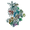
+ Open data
- Basic information
| Entry | Database: PDB / ID: 8jsg | ||||||
|---|---|---|---|---|---|---|---|
| Title | Structure of the 30S-IF3 complex from Escherichia coli | ||||||
 Components Components |
| ||||||
 Keywords Keywords | TRANSLATION / Ribosome / 30S / IF3 / Translation initiation | ||||||
| Function / homology |  Function and homology information Function and homology informationribosome disassembly / ornithine decarboxylase inhibitor activity / transcription antitermination factor activity, RNA binding / misfolded RNA binding / Group I intron splicing / RNA folding / four-way junction DNA binding / negative regulation of translational initiation / regulation of mRNA stability / translation initiation factor activity ...ribosome disassembly / ornithine decarboxylase inhibitor activity / transcription antitermination factor activity, RNA binding / misfolded RNA binding / Group I intron splicing / RNA folding / four-way junction DNA binding / negative regulation of translational initiation / regulation of mRNA stability / translation initiation factor activity / mRNA regulatory element binding translation repressor activity / positive regulation of RNA splicing / transcription elongation factor complex / regulation of DNA-templated transcription elongation / DNA endonuclease activity / transcription antitermination / DNA-templated transcription termination / maintenance of translational fidelity / mRNA 5'-UTR binding / ribosomal small subunit biogenesis / small ribosomal subunit rRNA binding / ribosome biogenesis / ribosome binding / regulation of translation / ribosomal small subunit assembly / small ribosomal subunit / cytosolic small ribosomal subunit / cytoplasmic translation / tRNA binding / molecular adaptor activity / negative regulation of translation / rRNA binding / ribosome / structural constituent of ribosome / translation / response to antibiotic / mRNA binding / RNA binding / zinc ion binding / membrane / cytosol / cytoplasm Similarity search - Function | ||||||
| Biological species |  | ||||||
| Method | ELECTRON MICROSCOPY / single particle reconstruction / cryo EM / Resolution: 4.6 Å | ||||||
 Authors Authors | Uday, A.B. / Mishra, R.K. / Hussain, T. | ||||||
| Funding support |  United Kingdom, 1items United Kingdom, 1items
| ||||||
 Citation Citation |  Journal: Proteins / Year: 2023 Journal: Proteins / Year: 2023Title: Initiation factor 3 bound to the 30S ribosomal subunit in an initial step of translation. Authors: Adwaith B Uday / Rishi Kumar Mishra / Tanweer Hussain /  Abstract: Bacterial ribosomes require three initiation factors IF1, IF2, and IF3 during the initial steps of translation. These IFs ensure correct base pairing of the initiator tRNA anticodon with the start ...Bacterial ribosomes require three initiation factors IF1, IF2, and IF3 during the initial steps of translation. These IFs ensure correct base pairing of the initiator tRNA anticodon with the start codon in the mRNA located at the P-site of the 30S ribosomal subunit. IF3 is one of the first IFs to bind to the 30S and plays a crucial role in the selection of the correct start codon and codon: anticodon base pairing. IF3 also prevents the premature association of the 50S subunit of ribosomes and aids in ribosome recycling. IF3 is reported to change binding sites and conformation to ensure translation initiation fidelity. A recent study suggested an initial binding of IF3 CTD away from the P-site and that IF1 and IF2 promote the movement of CTD to the P-site and concomitant movement of NTD. Hence, to visualize the position of IF3 in the absence of any other IFs, we determined cryo-EM structure of the 30S-IF3 complex. The map shows that IF3 is present in an extended conformation with CTD present at the P-site and NTD near the platform even in the absence of IF1 and IF2. Hence, IF3 CTD binds at the P-site and moves away during the accommodation of the initiator tRNA at the P-site in the later steps of translation initiation. Overall, we report the structure of 30S-IF3 which demystifies the starting binding site and conformation of IF3 on the 30S ribosomal subunit. | ||||||
| History |
|
- Structure visualization
| Structure viewer | Molecule:  Molmil Molmil Jmol/JSmol Jmol/JSmol |
|---|
- Downloads & links
- Download
| PDBx/mmCIF format |  8jsg.cif.gz 8jsg.cif.gz | 1.4 MB | Display |  PDBx/mmCIF format PDBx/mmCIF format |
|---|---|---|---|---|
| PDB format |  pdb8jsg.ent.gz pdb8jsg.ent.gz | 915.7 KB | Display |  PDB format PDB format |
| PDBx/mmJSON format |  8jsg.json.gz 8jsg.json.gz | Tree view |  PDBx/mmJSON format PDBx/mmJSON format | |
| Others |  Other downloads Other downloads |
-Validation report
| Summary document |  8jsg_validation.pdf.gz 8jsg_validation.pdf.gz | 1.3 MB | Display |  wwPDB validaton report wwPDB validaton report |
|---|---|---|---|---|
| Full document |  8jsg_full_validation.pdf.gz 8jsg_full_validation.pdf.gz | 1.3 MB | Display | |
| Data in XML |  8jsg_validation.xml.gz 8jsg_validation.xml.gz | 90.6 KB | Display | |
| Data in CIF |  8jsg_validation.cif.gz 8jsg_validation.cif.gz | 159.7 KB | Display | |
| Arichive directory |  https://data.pdbj.org/pub/pdb/validation_reports/js/8jsg https://data.pdbj.org/pub/pdb/validation_reports/js/8jsg ftp://data.pdbj.org/pub/pdb/validation_reports/js/8jsg ftp://data.pdbj.org/pub/pdb/validation_reports/js/8jsg | HTTPS FTP |
-Related structure data
| Related structure data |  36619MC  8jshC M: map data used to model this data C: citing same article ( |
|---|---|
| Similar structure data | Similarity search - Function & homology  F&H Search F&H Search |
- Links
Links
- Assembly
| Deposited unit | 
|
|---|---|
| 1 |
|
- Components
-Small ribosomal subunit protein ... , 19 types, 19 molecules 123Pklnpqtuhmorswzj
| #1: Protein | Mass: 8746.147 Da / Num. of mol.: 1 / Source method: isolated from a natural source / Source: (natural)  |
|---|---|
| #2: Protein | Mass: 6263.326 Da / Num. of mol.: 1 / Source method: isolated from a natural source / Source: (natural)  |
| #3: Protein | Mass: 9577.268 Da / Num. of mol.: 1 / Source method: isolated from a natural source / Source: (natural)  |
| #5: Protein | Mass: 9480.138 Da / Num. of mol.: 1 / Source method: isolated from a natural source / Source: (natural)  |
| #7: Protein | Mass: 16641.217 Da / Num. of mol.: 1 / Source method: isolated from a natural source / Source: (natural)  |
| #8: Protein | Mass: 23383.002 Da / Num. of mol.: 1 / Source method: isolated from a natural source / Source: (natural)  |
| #9: Protein | Mass: 11669.371 Da / Num. of mol.: 1 / Source method: isolated from a natural source / Source: (natural)  |
| #10: Protein | Mass: 14015.361 Da / Num. of mol.: 1 / Source method: isolated from a natural source / Source: (natural)  |
| #11: Protein | Mass: 13739.778 Da / Num. of mol.: 1 / Source method: isolated from a natural source / Source: (natural)  |
| #12: Protein | Mass: 13636.961 Da / Num. of mol.: 1 / Source method: isolated from a natural source / Source: (natural)  |
| #13: Protein | Mass: 10159.621 Da / Num. of mol.: 1 / Source method: isolated from a natural source / Source: (natural)  |
| #15: Protein | Mass: 23078.785 Da / Num. of mol.: 1 / Source method: isolated from a natural source / Source: (natural)  |
| #16: Protein | Mass: 16861.523 Da / Num. of mol.: 1 / Source method: isolated from a natural source / Source: (natural)  |
| #17: Protein | Mass: 14755.074 Da / Num. of mol.: 1 / Source method: isolated from a natural source / Source: (natural)  |
| #18: Protein | Mass: 11698.545 Da / Num. of mol.: 1 / Source method: isolated from a natural source / Source: (natural)  |
| #19: Protein | Mass: 12625.753 Da / Num. of mol.: 1 / Source method: isolated from a natural source / Source: (natural)  |
| #20: Protein | Mass: 11475.364 Da / Num. of mol.: 1 / Source method: isolated from a natural source / Source: (natural)  |
| #21: Protein | Mass: 9154.740 Da / Num. of mol.: 1 / Source method: isolated from a natural source / Source: (natural)  |
| #22: Protein | Mass: 25015.816 Da / Num. of mol.: 1 / Source method: isolated from a natural source / Source: (natural)  |
-Protein , 2 types, 2 molecules Ay
| #4: Protein | Mass: 20600.994 Da / Num. of mol.: 1 Source method: isolated from a genetically manipulated source Source: (gene. exp.)   |
|---|---|
| #14: Protein | Mass: 9207.572 Da / Num. of mol.: 1 / Source method: isolated from a natural source / Source: (natural)  |
-RNA chain , 1 types, 1 molecules g
| #6: RNA chain | Mass: 499077.625 Da / Num. of mol.: 1 / Source method: isolated from a natural source / Source: (natural)  |
|---|
-Experimental details
-Experiment
| Experiment | Method: ELECTRON MICROSCOPY |
|---|---|
| EM experiment | Aggregation state: PARTICLE / 3D reconstruction method: single particle reconstruction |
- Sample preparation
| Component |
| ||||||||||||||||||||||||||||||
|---|---|---|---|---|---|---|---|---|---|---|---|---|---|---|---|---|---|---|---|---|---|---|---|---|---|---|---|---|---|---|---|
| Molecular weight |
| ||||||||||||||||||||||||||||||
| Source (natural) |
| ||||||||||||||||||||||||||||||
| Source (recombinant) | Organism:  | ||||||||||||||||||||||||||||||
| Buffer solution | pH: 7.5 | ||||||||||||||||||||||||||||||
| Buffer component |
| ||||||||||||||||||||||||||||||
| Specimen | Embedding applied: NO / Shadowing applied: NO / Staining applied: NO / Vitrification applied: YES | ||||||||||||||||||||||||||||||
| Specimen support | Grid material: COPPER / Grid type: Quantifoil R2/2 | ||||||||||||||||||||||||||||||
| Vitrification | Instrument: FEI VITROBOT MARK IV / Cryogen name: ETHANE / Humidity: 100 % / Chamber temperature: 277 K |
- Electron microscopy imaging
| Experimental equipment |  Model: Talos Arctica / Image courtesy: FEI Company |
|---|---|
| Microscopy | Model: FEI TALOS ARCTICA |
| Electron gun | Electron source:  FIELD EMISSION GUN / Accelerating voltage: 200 kV / Illumination mode: OTHER FIELD EMISSION GUN / Accelerating voltage: 200 kV / Illumination mode: OTHER |
| Electron lens | Mode: OTHER / Nominal magnification: 45000 X / Nominal defocus max: 2500 nm / Nominal defocus min: 1500 nm / Cs: 2.7 mm |
| Specimen holder | Cryogen: NITROGEN |
| Image recording | Electron dose: 60 e/Å2 / Film or detector model: GATAN K2 SUMMIT (4k x 4k) / Num. of grids imaged: 2 / Num. of real images: 4564 |
- Processing
| EM software | Name: PHENIX / Version: 1.21rc1_4985 / Category: model refinement | ||||||||||||||||||||||||
|---|---|---|---|---|---|---|---|---|---|---|---|---|---|---|---|---|---|---|---|---|---|---|---|---|---|
| CTF correction | Type: PHASE FLIPPING AND AMPLITUDE CORRECTION | ||||||||||||||||||||||||
| 3D reconstruction | Resolution: 4.6 Å / Resolution method: FSC 0.143 CUT-OFF / Num. of particles: 145664 / Symmetry type: POINT | ||||||||||||||||||||||||
| Refinement | Cross valid method: NONE Stereochemistry target values: GeoStd + Monomer Library + CDL v1.2 | ||||||||||||||||||||||||
| Displacement parameters | Biso mean: 119.73 Å2 | ||||||||||||||||||||||||
| Refine LS restraints |
|
 Movie
Movie Controller


 PDBj
PDBj


