+ Open data
- Basic information
| Entry | Database: PDB / ID: 8jq9 | ||||||
|---|---|---|---|---|---|---|---|
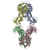
| Title | Structure of Gabija GajA | ||||||
 Components Components | Endonuclease GajA | ||||||
 Keywords Keywords | IMMUNE SYSTEM / Anti-Phage | ||||||
| Function / homology |  Function and homology information Function and homology informationendonuclease activity / defense response to virus / Hydrolases; Acting on ester bonds / metal ion binding Similarity search - Function | ||||||
| Biological species |  | ||||||
| Method | ELECTRON MICROSCOPY / single particle reconstruction / cryo EM / Resolution: 2.66 Å | ||||||
 Authors Authors | Li, J. / Wang, Z. / Wang, L. | ||||||
| Funding support |  China, 1items China, 1items
| ||||||
 Citation Citation |  Journal: Nature / Year: 2024 Journal: Nature / Year: 2024Title: Structures and activation mechanism of the Gabija anti-phage system. Authors: Jing Li / Rui Cheng / Zhiming Wang / Wuliu Yuan / Jun Xiao / Xinyuan Zhao / Xinran Du / Shiyu Xia / Lianrong Wang / Bin Zhu / Longfei Wang /   Abstract: Prokaryotes have evolved intricate innate immune systems against phage infection. Gabija is a highly widespread prokaryotic defence system that consists of two components, GajA and GajB. GajA ...Prokaryotes have evolved intricate innate immune systems against phage infection. Gabija is a highly widespread prokaryotic defence system that consists of two components, GajA and GajB. GajA functions as a DNA endonuclease that is inactive in the presence of ATP. Here, to explore how the Gabija system is activated for anti-phage defence, we report its cryo-electron microscopy structures in five states, including apo GajA, GajA in complex with DNA, GajA bound by ATP, apo GajA-GajB, and GajA-GajB in complex with ATP and Mg. GajA is a rhombus-shaped tetramer with its ATPase domain clustered at the centre and the topoisomerase-primase (Toprim) domain located peripherally. ATP binding at the ATPase domain stabilizes the insertion region within the ATPase domain, keeping the Toprim domain in a closed state. Upon ATP depletion by phages, the Toprim domain opens to bind and cleave the DNA substrate. GajB, which docks on GajA, is activated by the cleaved DNA, ultimately leading to prokaryotic cell death. Our study presents a mechanistic landscape of Gabija activation. | ||||||
| History |
|
- Structure visualization
| Structure viewer | Molecule:  Molmil Molmil Jmol/JSmol Jmol/JSmol |
|---|
- Downloads & links
- Download
| PDBx/mmCIF format |  8jq9.cif.gz 8jq9.cif.gz | 452.6 KB | Display |  PDBx/mmCIF format PDBx/mmCIF format |
|---|---|---|---|---|
| PDB format |  pdb8jq9.ent.gz pdb8jq9.ent.gz | 298.9 KB | Display |  PDB format PDB format |
| PDBx/mmJSON format |  8jq9.json.gz 8jq9.json.gz | Tree view |  PDBx/mmJSON format PDBx/mmJSON format | |
| Others |  Other downloads Other downloads |
-Validation report
| Summary document |  8jq9_validation.pdf.gz 8jq9_validation.pdf.gz | 373.5 KB | Display |  wwPDB validaton report wwPDB validaton report |
|---|---|---|---|---|
| Full document |  8jq9_full_validation.pdf.gz 8jq9_full_validation.pdf.gz | 381.4 KB | Display | |
| Data in XML |  8jq9_validation.xml.gz 8jq9_validation.xml.gz | 32.9 KB | Display | |
| Data in CIF |  8jq9_validation.cif.gz 8jq9_validation.cif.gz | 50.7 KB | Display | |
| Arichive directory |  https://data.pdbj.org/pub/pdb/validation_reports/jq/8jq9 https://data.pdbj.org/pub/pdb/validation_reports/jq/8jq9 ftp://data.pdbj.org/pub/pdb/validation_reports/jq/8jq9 ftp://data.pdbj.org/pub/pdb/validation_reports/jq/8jq9 | HTTPS FTP |
-Related structure data
| Related structure data |  36541MC  8jqbC  8jqcC  8wy4C  8wy5C  8x51C  8x5iC  8x5nC M: map data used to model this data C: citing same article ( |
|---|---|
| Similar structure data | Similarity search - Function & homology  F&H Search F&H Search |
- Links
Links
- Assembly
| Deposited unit | 
|
|---|---|
| 1 |
|
- Components
| #1: Protein | Mass: 67079.469 Da / Num. of mol.: 4 Source method: isolated from a genetically manipulated source Source: (gene. exp.)  Gene: gajA, IIE_04982 / Production host:  References: UniProt: J8H9C1, Hydrolases; Acting on ester bonds Has protein modification | N | |
|---|
-Experimental details
-Experiment
| Experiment | Method: ELECTRON MICROSCOPY |
|---|---|
| EM experiment | Aggregation state: PARTICLE / 3D reconstruction method: single particle reconstruction |
- Sample preparation
| Component | Name: GajA / Type: COMPLEX / Entity ID: all / Source: RECOMBINANT |
|---|---|
| Molecular weight | Value: 0.276 MDa / Experimental value: NO |
| Source (natural) | Organism:  |
| Source (recombinant) | Organism:  |
| Buffer solution | pH: 8 |
| Specimen | Embedding applied: NO / Shadowing applied: NO / Staining applied: NO / Vitrification applied: YES |
| Vitrification | Instrument: FEI VITROBOT MARK IV / Cryogen name: ETHANE / Humidity: 100 % / Chamber temperature: 8 K |
- Electron microscopy imaging
| Experimental equipment |  Model: Titan Krios / Image courtesy: FEI Company |
|---|---|
| Microscopy | Model: FEI TITAN KRIOS |
| Electron gun | Electron source:  FIELD EMISSION GUN / Accelerating voltage: 300 kV / Illumination mode: FLOOD BEAM FIELD EMISSION GUN / Accelerating voltage: 300 kV / Illumination mode: FLOOD BEAM |
| Electron lens | Mode: BRIGHT FIELD / Nominal defocus max: 1800 nm / Nominal defocus min: 1600 nm |
| Image recording | Electron dose: 50 e/Å2 / Film or detector model: GATAN K3 (6k x 4k) |
- Processing
| CTF correction | Type: PHASE FLIPPING AND AMPLITUDE CORRECTION | ||||||||||||||||||||||||
|---|---|---|---|---|---|---|---|---|---|---|---|---|---|---|---|---|---|---|---|---|---|---|---|---|---|
| 3D reconstruction | Resolution: 2.66 Å / Resolution method: FSC 0.143 CUT-OFF / Num. of particles: 3023069 / Symmetry type: POINT | ||||||||||||||||||||||||
| Atomic model building | Protocol: AB INITIO MODEL / Space: REAL | ||||||||||||||||||||||||
| Refinement | Cross valid method: NONE Stereochemistry target values: GeoStd + Monomer Library + CDL v1.2 | ||||||||||||||||||||||||
| Displacement parameters | Biso mean: 48.19 Å2 | ||||||||||||||||||||||||
| Refine LS restraints |
|
 Movie
Movie Controller


 PDBj
PDBj