 Movie
Movie Controller


+ Open data

- Basic information
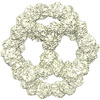
| Entry | Database: PDB / ID: 8jgc | ||||||
|---|---|---|---|---|---|---|---|
| Title | Cryo-EM structure of Mi3 fused with LOV2 | ||||||
 Components Components | LOV domain-containing protein,2-dehydro-3-deoxyphosphogluconate aldolase/4-hydroxy-2-oxoglutarate aldolase | ||||||
 Keywords Keywords | LYASE / protein cage Scaffold | ||||||
| Function / homology |  Function and homology information Function and homology informationresponse to blue light / photoreceptor activity / non-specific serine/threonine protein kinase / lyase activity / protein kinase activity / ATP binding Similarity search - Function | ||||||
| Biological species |    Thermotoga maritima (bacteria) Thermotoga maritima (bacteria) | ||||||
| Method | ELECTRON MICROSCOPY / single particle reconstruction / cryo EM / Resolution: 3.44 Å | ||||||
 Authors Authors | Zhang, H.W. / Kang, W. / Xue, C. | ||||||
| Funding support |  China, 1items China, 1items
| ||||||
 Citation Citation | Journal: J Am Chem Soc / Year: 2024 Title: Dynamic Metabolons Using Stimuli-Responsive Protein Cages. Authors: Wei Kang / Xiao Ma / Huawei Zhang / Juncai Ma / Chunxue Liu / Jiani Li / Hanhan Guo / Daping Wang / Rui Wang / Bo Li / Chuang Xue /  Abstract: Naturally evolved metabolons have the ability to assemble and disassemble in response to environmental stimuli, allowing for the rapid reorganization of chemical reactions in living cells to meet ...Naturally evolved metabolons have the ability to assemble and disassemble in response to environmental stimuli, allowing for the rapid reorganization of chemical reactions in living cells to meet changing cellular needs. However, replicating such capability in synthetic metabolons remains a challenge due to our limited understanding of the mechanisms by which the assembly and disassembly of such naturally occurring multienzyme complexes are controlled. Here, we report the synthesis of chemical- and light-responsive protein cages for assembling synthetic metabolons, enabling the dynamic regulation of enzymatic reactions in living cells. Particularly, a chemically responsive domain was fused to a self-assembled protein cage subunit, generating engineered protein cages capable of displaying proteins containing cognate interaction domains on their surfaces in response to small molecular cues. Chemical-induced colocalization of sequential enzymes on protein cages enhances the specificity of the branched deoxyviolacein biosynthetic reactions by 2.6-fold. Further, by replacing the chemical-inducible domain with a light-inducible dimerization domain, we created an optogenetic protein cage capable of reversibly recruiting and releasing targeted proteins onto and from the exterior of the protein cages in tens of seconds by on-off of blue light. Tethering the optogenetic protein cages to membranes enables the formation of light-switchable, membrane-bound metabolons, which can repeatably recruit-release enzymes, leading to the manipulation of substrate utilization across membranes on demand. Our work demonstrates a powerful and versatile strategy for constructing dynamic metabolons in engineered living cells for efficient and controllable biocatalysis. | ||||||
| History |
|
- Structure visualization
| Structure viewer | Molecule: MolmilJmol/JSmol |
|---|
- Downloads & links
-Download
| PDBx/mmCIF format | 8jgc.cif.gz | 50.3 KB | Display | PDBx/mmCIF format |
|---|---|---|---|---|
| PDB format | pdb8jgc.ent.gz | 32.2 KB | Display | PDB format |
| PDBx/mmJSON format | 8jgc.json.gz | Tree view | PDBx/mmJSON format | |
| Others |  Other downloads Other downloads |
-Validation report
| Summary document | 8jgc_validation.pdf.gz | 1.3 MB | Display | wwPDB validaton report |
|---|---|---|---|---|
| Full document | 8jgc_full_validation.pdf.gz | 1.3 MB | Display | |
| Data in XML | 8jgc_validation.xml.gz | 34.3 KB | Display | |
| Data in CIF | 8jgc_validation.cif.gz | 46.4 KB | Display | |
| Arichive directory | https://data.pdbj.org/pub/pdb/validation_reports/jg/8jgcftp://data.pdbj.org/pub/pdb/validation_reports/jg/8jgc | HTTPS FTP |
-Related structure data
| Related structure data | 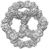 36230MC  8jgaC M: map data used to model this data C: citing same article ( |
|---|---|
| Similar structure data |
-Links
 PDBj
PDBj


- Assembly
| Deposited unit | 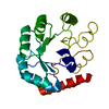
|
|---|---|
| 1 |
|
-Components
| #1: Protein | Mass: 41424.469 Da / Num. of mol.: 1 Source method: isolated from a genetically manipulated source Source: (gene. exp.) Thermotoga maritima (strain ATCC 43589 / DSM 3109 / JCM 10099 / NBRC 100826 / MSB8) (bacteria)Gene: TM_0066 / Production host: |
|---|
-Experimental details
-Experiment
| Experiment | Method: ELECTRON MICROSCOPY |
|---|---|
| EM experiment | Aggregation state: PARTICLE / 3D reconstruction method: single particle reconstruction |
- Sample preparation
| Component | Name: LOV2-Mi3 / Type: COMPLEX / Entity ID: all / Source: MULTIPLE SOURCES |
|---|---|
| Source (natural) | Organism: Thermotoga maritima (bacteria) |
| Source (recombinant) | Organism: |
| Buffer solution | pH: 7.8 |
| Specimen | Embedding applied: NO / Shadowing applied: NO / Staining applied: NO / Vitrification applied: YES |
| Vitrification | Cryogen name: ETHANE |
- Electron microscopy imaging
| Experimental equipment |  Model: Titan Krios / Image courtesy: FEI Company |
|---|---|
| Microscopy | Model: FEI TITAN KRIOS |
| Electron gun | Electron source:  FIELD EMISSION GUN / Accelerating voltage: 300 kV / Illumination mode: FLOOD BEAM FIELD EMISSION GUN / Accelerating voltage: 300 kV / Illumination mode: FLOOD BEAM |
| Electron lens | Mode: BRIGHT FIELD / Nominal defocus max: 2500 nm / Nominal defocus min: 1500 nm |
| Image recording | Electron dose: 50 e/Å2 / Film or detector model: GATAN K3 BIOQUANTUM (6k x 4k) |
- Processing
| EM software | Name: PHENIX / Version: dev_3951: / Category: model refinement | ||||||||||||||||||||||||
|---|---|---|---|---|---|---|---|---|---|---|---|---|---|---|---|---|---|---|---|---|---|---|---|---|---|
| CTF correction | Type: PHASE FLIPPING AND AMPLITUDE CORRECTION | ||||||||||||||||||||||||
| 3D reconstruction | Resolution: 3.44 Å / Resolution method: FSC 0.143 CUT-OFF / Num. of particles: 85541 / Symmetry type: POINT | ||||||||||||||||||||||||
| Refine LS restraints |
|
