 Movie
Movie Controller


+ Open data

- Basic information
| Entry | Database: PDB / ID: 8hwb | ||||||
|---|---|---|---|---|---|---|---|
| Title | D5 ATP-ADP-Apo-ssDNA IS2 | ||||||
 Components Components |
| ||||||
 Keywords Keywords | VIRAL PROTEIN / MPVX | ||||||
| Function / homology |  Function and homology information Function and homology information | ||||||
| Biological species |  Monkeypox virus Monkeypox virus | ||||||
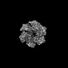
| Method | ELECTRON MICROSCOPY / single particle reconstruction / cryo EM / Resolution: 3.9 Å | ||||||
 Authors Authors | Li, Y.N. / Zhu, J. / Guo, Y.Y. / Yan, R.H. | ||||||
| Funding support |  China, 1items China, 1items
| ||||||
 Citation Citation | Journal: Nat Struct Mol Biol / Year: 2024 Title: Structural insight into the assembly and working mechanism of helicase-primase D5 from Mpox virus. Authors: Yaning Li / Jing Zhu / Yingying Guo / Renhong Yan / Abstract: The Mpox pandemic, caused by the Mpox virus (or monkeypox virus, MPXV), has gained global attention. The D5 protein, a putative helicase-primase found in MPXV, plays a vital role in viral replication ...The Mpox pandemic, caused by the Mpox virus (or monkeypox virus, MPXV), has gained global attention. The D5 protein, a putative helicase-primase found in MPXV, plays a vital role in viral replication and genome uncoating. Here we determined multiple cryo-EM structures of full-length hexameric D5 in diverse states. These states were captured during ATP hydrolysis while moving along the single-stranded DNA (ssDNA) track. Through comprehensive structural analysis combined with the helicase activity system, we revealed that when the primase domain is truncated or the interaction between the primase and helicase domains is disrupted, the double-stranded DNA (dsDNA) unwinds into ssDNA, suggesting a critical regulatory role of the primase domain. Two transition states bound with ssDNA substrate during unwinding reveals that two ATP molecules were consumed to drive DNA moving forward two nucleotides. Collectively, our findings shed light on the molecular mechanism that links ATP hydrolysis to the DNA unwinding in poxviruses. | ||||||
| History |
|
- Structure visualization
| Structure viewer | Molecule: MolmilJmol/JSmol |
|---|
- Downloads & links
-Download
| PDBx/mmCIF format | 8hwb.cif.gz | 590.6 KB | Display | PDBx/mmCIF format |
|---|---|---|---|---|
| PDB format | pdb8hwb.ent.gz | 471.2 KB | Display | PDB format |
| PDBx/mmJSON format | 8hwb.json.gz | Tree view | PDBx/mmJSON format | |
| Others |  Other downloads Other downloads |
-Validation report
| Arichive directory | https://data.pdbj.org/pub/pdb/validation_reports/hw/8hwbftp://data.pdbj.org/pub/pdb/validation_reports/hw/8hwb | HTTPS FTP |
|---|
-Related structure data
| Related structure data |  35052MC  8hwaC  8hwcC  8hwdC 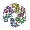 8hweC  8hwfC 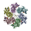 8hwgC  8hwhC M: map data used to model this data C: citing same article ( |
|---|---|
| Similar structure data |


-Links
 PDBj
PDBj

- Assembly
| Deposited unit | 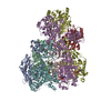
|
|---|---|
| 1 |
|
-Components
| #1: Protein | Mass: 90476.344 Da / Num. of mol.: 7 Source method: isolated from a genetically manipulated source Source: (gene. exp.) Monkeypox virusGene: E5R, MPXV-COP-096, MPXV-M2940_FCT-100, MPXV-M2957_Lagos-100, MPXV-M3021_Delta-100, MPXV-M5320_M15_Bayelsa-093, MPXV-Nig_SEV71_2_82-095, MPXV-PCH-097, MPXV-Singapore-100, MPXV-SL-096, MPXV-UK_P1- ...Gene: E5R, MPXV-COP-096, MPXV-M2940_FCT-100, MPXV-M2957_Lagos-100, MPXV-M3021_Delta-100, MPXV-M5320_M15_Bayelsa-093, MPXV-Nig_SEV71_2_82-095, MPXV-PCH-097, MPXV-Singapore-100, MPXV-SL-096, MPXV-UK_P1-100, MPXV-UK_P2-100, MPXV-UK_P3-100, MPXV-USA2003_099_GR-100, MPXV-USA2003_206_DM-100, MPXV-USA2003_223_RS-100, MPXV-UTC-091, MPXV-W_Nigeria-095, MPXV-WRAIR096, MPXV298464_082, MPXV_LIB1970_184_107, MPXV_USA2003_039_107, MPXV_USA2003_044_107, PDLMKLCO_00105 Production host:  Homo sapiens (human) / References: UniProt: Q5IXS3 Homo sapiens (human) / References: UniProt: Q5IXS3#2: DNA chain | | Mass: 1780.199 Da / Num. of mol.: 1 / Source method: obtained synthetically / Source: (synth.) Monkeypox virus#3: Chemical | ChemComp-ATP /   Mass: 507.181 Da / Num. of mol.: 5 / Source method: obtained synthetically / Formula: C10H16N5O13P3 / Feature type: SUBJECT OF INVESTIGATION / Comment: ATP, energy-carrying molecule*YM Mass: 507.181 Da / Num. of mol.: 5 / Source method: obtained synthetically / Formula: C10H16N5O13P3 / Feature type: SUBJECT OF INVESTIGATION / Comment: ATP, energy-carrying molecule*YM#4: Chemical | ChemComp-MG /   Mass: 24.305 Da / Num. of mol.: 4 / Source method: obtained synthetically / Formula: Mg / Feature type: SUBJECT OF INVESTIGATION Mass: 24.305 Da / Num. of mol.: 4 / Source method: obtained synthetically / Formula: Mg / Feature type: SUBJECT OF INVESTIGATION#5: Chemical | ChemComp-ADP / |   Mass: 427.201 Da / Num. of mol.: 1 / Source method: obtained synthetically / Formula: C10H15N5O10P2 / Feature type: SUBJECT OF INVESTIGATION / Comment: ADP, energy-carrying molecule*YM Mass: 427.201 Da / Num. of mol.: 1 / Source method: obtained synthetically / Formula: C10H15N5O10P2 / Feature type: SUBJECT OF INVESTIGATION / Comment: ADP, energy-carrying molecule*YMHas ligand of interest | Y | |
|---|
-Experimental details
-Experiment
| Experiment | Method: ELECTRON MICROSCOPY |
|---|---|
| EM experiment | Aggregation state: PARTICLE / 3D reconstruction method: single particle reconstruction |
- Sample preparation
| Component | Name: D5 ATP-ADP-Apo-ssDNA IS2 / Type: COMPLEX / Entity ID: #1-#2 / Source: MULTIPLE SOURCES |
|---|---|
| Source (natural) | Organism: Monkeypox virus |
| Source (recombinant) | Organism: Homo sapiens (human) |
| Buffer solution | pH: 7.5 |
| Specimen | Embedding applied: NO / Shadowing applied: NO / Staining applied: NO / Vitrification applied: YES |
| Vitrification | Cryogen name: ETHANE |
- Electron microscopy imaging
| Experimental equipment |  Model: Titan Krios / Image courtesy: FEI Company |
|---|---|
| Microscopy | Model: FEI TITAN KRIOS |
| Electron gun | Electron source:  FIELD EMISSION GUN / Accelerating voltage: 300 kV / Illumination mode: FLOOD BEAM FIELD EMISSION GUN / Accelerating voltage: 300 kV / Illumination mode: FLOOD BEAM |
| Electron lens | Mode: BRIGHT FIELD / Nominal defocus max: 2200 nm / Nominal defocus min: 1200 nm / Alignment procedure: COMA FREE |
| Specimen holder | Cryogen: NITROGEN / Specimen holder model: FEI TITAN KRIOS AUTOGRID HOLDER |
| Image recording | Electron dose: 50 e/Å2 / Film or detector model: GATAN K3 BIOQUANTUM (6k x 4k) |
- Processing
| EM software | Name: RELION / Version: 3.0.6 / Category: 3D reconstruction |
|---|---|
| CTF correction | Type: PHASE FLIPPING AND AMPLITUDE CORRECTION |
| 3D reconstruction | Resolution: 3.9 Å / Resolution method: FSC 0.143 CUT-OFF / Num. of particles: 67228 / Symmetry type: POINT |
