+ Open data
- Basic information
| Entry | Database: PDB / ID: 8hbw | ||||||||||||||||||
|---|---|---|---|---|---|---|---|---|---|---|---|---|---|---|---|---|---|---|---|
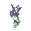
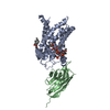
| Title | Structure of human UCP1 in the ATP-bound state | ||||||||||||||||||
 Components Components |
| ||||||||||||||||||
 Keywords Keywords | MEMBRANE PROTEIN / UCP1 / SLC25A7 / thermogenin / SLC25 | ||||||||||||||||||
| Function / homology |  Function and homology information Function and homology informationpurine ribonucleotide binding / cellular response to dehydroepiandrosterone / Mitochondrial Uncoupling / The fatty acid cycling model / oxidative phosphorylation uncoupler activity / mitochondrial transmembrane transport / adaptive thermogenesis / cardiolipin binding / regulation of reactive oxygen species biosynthetic process / cellular response to cold ...purine ribonucleotide binding / cellular response to dehydroepiandrosterone / Mitochondrial Uncoupling / The fatty acid cycling model / oxidative phosphorylation uncoupler activity / mitochondrial transmembrane transport / adaptive thermogenesis / cardiolipin binding / regulation of reactive oxygen species biosynthetic process / cellular response to cold / cellular response to fatty acid / response to temperature stimulus / long-chain fatty acid binding / diet induced thermogenesis / proton transmembrane transporter activity / transmembrane transporter activity / brown fat cell differentiation / Transcriptional regulation of brown and beige adipocyte differentiation by EBF2 / cellular response to hormone stimulus / response to cold / proton transmembrane transport / response to nutrient levels / cellular response to reactive oxygen species / GDP binding / positive regulation of cold-induced thermogenesis / mitochondrial inner membrane / GTP binding / regulation of transcription by RNA polymerase II / mitochondrion Similarity search - Function | ||||||||||||||||||
| Biological species |  Homo sapiens (human) Homo sapiens (human)synthetic construct (others) | ||||||||||||||||||
| Method | ELECTRON MICROSCOPY / single particle reconstruction / cryo EM / Resolution: 2.57 Å | ||||||||||||||||||
 Authors Authors | Chen, L. / Kang, Y. | ||||||||||||||||||
| Funding support |  China, 5items China, 5items
| ||||||||||||||||||
 Citation Citation |  Journal: Nature / Year: 2023 Journal: Nature / Year: 2023Title: Structural basis for the binding of DNP and purine nucleotides onto UCP1. Authors: Yunlu Kang / Lei Chen /  Abstract: Uncoupling protein 1 (UCP1) conducts protons through the inner mitochondrial membrane to uncouple mitochondrial respiration from ATP production, thereby converting the electrochemical gradient of ...Uncoupling protein 1 (UCP1) conducts protons through the inner mitochondrial membrane to uncouple mitochondrial respiration from ATP production, thereby converting the electrochemical gradient of protons into heat. The activity of UCP1 is activated by endogenous fatty acids and synthetic small molecules, such as 2,4-dinitrophenol (DNP), and is inhibited by purine nucleotides, such as ATP. However, the mechanism by which UCP1 binds to these ligands remains unknown. Here we present the structures of human UCP1 in the nucleotide-free state, the DNP-bound state and the ATP-bound state. The structures show that the central cavity of UCP1 is open to the cytosolic side. DNP binds inside the cavity, making contact with transmembrane helix 2 (TM2) and TM6. ATP binds in the same cavity and induces conformational changes in TM2, together with the inward bending of TM1, TM4, TM5 and TM6 of UCP1, resulting in a more compact structure of UCP1. The binding site of ATP overlaps with that of DNP, suggesting that ATP competitively blocks the functional engagement of DNP, resulting in the inhibition of the proton-conducting activity of UCP1. | ||||||||||||||||||
| History |
|
- Structure visualization
| Structure viewer | Molecule:  Molmil Molmil Jmol/JSmol Jmol/JSmol |
|---|
- Downloads & links
- Download
| PDBx/mmCIF format |  8hbw.cif.gz 8hbw.cif.gz | 93.5 KB | Display |  PDBx/mmCIF format PDBx/mmCIF format |
|---|---|---|---|---|
| PDB format |  pdb8hbw.ent.gz pdb8hbw.ent.gz | 63.9 KB | Display |  PDB format PDB format |
| PDBx/mmJSON format |  8hbw.json.gz 8hbw.json.gz | Tree view |  PDBx/mmJSON format PDBx/mmJSON format | |
| Others |  Other downloads Other downloads |
-Validation report
| Summary document |  8hbw_validation.pdf.gz 8hbw_validation.pdf.gz | 590 KB | Display |  wwPDB validaton report wwPDB validaton report |
|---|---|---|---|---|
| Full document |  8hbw_full_validation.pdf.gz 8hbw_full_validation.pdf.gz | 598.8 KB | Display | |
| Data in XML |  8hbw_validation.xml.gz 8hbw_validation.xml.gz | 11.1 KB | Display | |
| Data in CIF |  8hbw_validation.cif.gz 8hbw_validation.cif.gz | 15.8 KB | Display | |
| Arichive directory |  https://data.pdbj.org/pub/pdb/validation_reports/hb/8hbw https://data.pdbj.org/pub/pdb/validation_reports/hb/8hbw ftp://data.pdbj.org/pub/pdb/validation_reports/hb/8hbw ftp://data.pdbj.org/pub/pdb/validation_reports/hb/8hbw | HTTPS FTP |
-Related structure data
| Related structure data |  34645MC  8hbvC  8j1nC M: map data used to model this data C: citing same article ( |
|---|---|
| Similar structure data | Similarity search - Function & homology  F&H Search F&H Search |
- Links
Links
- Assembly
| Deposited unit | 
|
|---|---|
| 1 |
|
- Components
| #1: Protein | Mass: 39468.367 Da / Num. of mol.: 1 Source method: isolated from a genetically manipulated source Source: (gene. exp.)  Homo sapiens (human) / Gene: UCP1, SLC25A7, UCP / Production host: Homo sapiens (human) / Gene: UCP1, SLC25A7, UCP / Production host:  Homo sapiens (human) / References: UniProt: P25874 Homo sapiens (human) / References: UniProt: P25874 | ||||
|---|---|---|---|---|---|
| #2: Antibody | Mass: 15004.661 Da / Num. of mol.: 1 / Source method: obtained synthetically / Source: (synth.) synthetic construct (others) | ||||
| #3: Chemical | ChemComp-ATP / | ||||
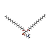
| #4: Chemical | ChemComp-CDL / | ||||
| #5: Chemical | | Has ligand of interest | Y | Has protein modification | Y | |
-Experimental details
-Experiment
| Experiment | Method: ELECTRON MICROSCOPY |
|---|---|
| EM experiment | Aggregation state: PARTICLE / 3D reconstruction method: single particle reconstruction |
- Sample preparation
| Component | Name: UCP1-sybody complex / Type: COMPLEX / Entity ID: #1-#2 / Source: MULTIPLE SOURCES |
|---|---|
| Source (natural) | Organism:  Homo sapiens (human) Homo sapiens (human) |
| Source (recombinant) | Organism:  Homo sapiens (human) Homo sapiens (human) |
| Buffer solution | pH: 7.5 |
| Specimen | Embedding applied: NO / Shadowing applied: NO / Staining applied: NO / Vitrification applied: YES |
| Vitrification | Cryogen name: ETHANE |
- Electron microscopy imaging
| Experimental equipment |  Model: Titan Krios / Image courtesy: FEI Company |
|---|---|
| Microscopy | Model: FEI TITAN KRIOS |
| Electron gun | Electron source:  FIELD EMISSION GUN / Accelerating voltage: 300 kV / Illumination mode: FLOOD BEAM FIELD EMISSION GUN / Accelerating voltage: 300 kV / Illumination mode: FLOOD BEAM |
| Electron lens | Mode: BRIGHT FIELD / Nominal defocus max: 1800 nm / Nominal defocus min: 1500 nm |
| Image recording | Electron dose: 52 e/Å2 / Film or detector model: GATAN K3 BIOQUANTUM (6k x 4k) |
- Processing
| CTF correction | Type: NONE |
|---|---|
| 3D reconstruction | Resolution: 2.57 Å / Resolution method: FSC 0.143 CUT-OFF / Num. of particles: 545929 / Symmetry type: POINT |
 Movie
Movie Controller


 PDBj
PDBj


