 Movie
Movie Controller


+ Open data

- Basic information
| Entry | Database: PDB / ID: 8gpj | ||||||
|---|---|---|---|---|---|---|---|
| Title | HIV-1 Env X16 UFO in complex with 8ANC195 Fab | ||||||
 Components Components |
| ||||||
 Keywords Keywords | VIRAL PROTEIN / Antibody / HIV-1 / Env protein / Complex | ||||||
| Function / homology | Immunoglobulins / Immunoglobulin-like / Sandwich / Mainly Beta Function and homology information Function and homology information | ||||||
| Biological species |  Homo sapiens (human) Homo sapiens (human) | ||||||
| Method | ELECTRON MICROSCOPY / single particle reconstruction / cryo EM / Resolution: 3.5 Å | ||||||
 Authors Authors | Niu, J. / Xu, Y.W. / Yang, B. | ||||||
| Funding support |  China, 1items China, 1items
| ||||||
 Citation Citation | Journal: Nat Commun / Year: 2023 Title: Structures and immune recognition of Env trimers from two Asia prevalent HIV-1 CRFs. Authors: Jun Niu / Qi Wang / Wenwen Zhao / Bing Meng / Youwei Xu / Xianfang Zhang / Yi Feng / Qilian Qi / Yanling Hao / Xuan Zhang / Ying Liu / Jiangchao Xiang / Yiming Shao / Bei Yang / Abstract: Structure-guided immunofocusing HIV-1 vaccine design entails a comprehensive understanding of Envs from diverse HIV-1 subtypes, including circulating recombinant forms (CRFs). Here, we present the ...Structure-guided immunofocusing HIV-1 vaccine design entails a comprehensive understanding of Envs from diverse HIV-1 subtypes, including circulating recombinant forms (CRFs). Here, we present the cryo-EM structures of Envs from two Asia prevalent CRFs (CRF01_AE and CRF07_BC) at 3.0 and 3.5 Å. We compare the structures and glycosylation patterns of Envs from different subtypes and perform cross-clade statistical analyses to reveal the unique features of CRF01_AE V1 region, which are associated with the resistance to certain bNAbs. We also solve a 4.1 Å cryo-EM structure of CRF01_AE Env in complex with F6, the first bNAb from CRF01_AE-infected individuals. F6 recognizes a gp120-gp41 spanning epitope to allosterically destabilize the Env trimer apex and weaken inter-protomer packing, which in turn hinders the receptor binding and induces Env trimer disassembly, demonstrating a dual mechanism of neutralization. These findings broaden our understanding of CRF Envs and shed lights on immunofocusing HIV-1 vaccine design. | ||||||
| History |
|
- Structure visualization
| Structure viewer | Molecule: MolmilJmol/JSmol |
|---|
- Downloads & links
-Download
| PDBx/mmCIF format | 8gpj.cif.gz | 494.6 KB | Display | PDBx/mmCIF format |
|---|---|---|---|---|
| PDB format | pdb8gpj.ent.gz | 386.6 KB | Display | PDB format |
| PDBx/mmJSON format | 8gpj.json.gz | Tree view | PDBx/mmJSON format | |
| Others |  Other downloads Other downloads |
-Validation report
| Arichive directory | https://data.pdbj.org/pub/pdb/validation_reports/gp/8gpjftp://data.pdbj.org/pub/pdb/validation_reports/gp/8gpj | HTTPS FTP |
|---|
-Related structure data
| Related structure data |  34194MC 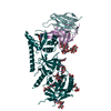 8gp5C  8gpgC  8gpiC 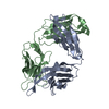 8gpkC C: citing same article ( M: map data used to model this data |
|---|---|
| Similar structure data |


-Links
 PDBj
PDBj


- Assembly
| Deposited unit | 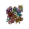
|
|---|---|
| 1 |
|
-Components
-Protein , 1 types, 6 molecules UVWXYZ
| #3: Protein | Mass: 69311.141 Da / Num. of mol.: 6 Source method: isolated from a genetically manipulated source Source: (gene. exp.) Homo sapiens (human) / Production host:   Cricetulus griseus (Chinese hamster) Cricetulus griseus (Chinese hamster) |
|---|
-Antibody , 2 types, 6 molecules ADHCEL
| #1: Antibody | Mass: 24923.986 Da / Num. of mol.: 3 Source method: isolated from a genetically manipulated source Source: (gene. exp.) Homo sapiens (human) / Production host:  Trichopalpus nigribasis (fry) Trichopalpus nigribasis (fry)#2: Antibody | Mass: 23401.984 Da / Num. of mol.: 3 Source method: isolated from a genetically manipulated source Source: (gene. exp.) Homo sapiens (human) / Production host: Trichopalpus nigribasis (fry) |
|---|
-Sugars , 7 types, 52 molecules 
| #4: Polysaccharide | beta-D-mannopyranose-(1-4)-2-acetamido-2-deoxy-beta-D-glucopyranose-(1-4)-2-acetamido-2-deoxy-beta- ...beta-D-mannopyranose-(1-4)-2-acetamido-2-deoxy-beta-D-glucopyranose-(1-4)-2-acetamido-2-deoxy-beta-D-glucopyranose Source method: isolated from a genetically manipulated source #5: Polysaccharide | 2-acetamido-2-deoxy-beta-D-glucopyranose-(1-4)-2-acetamido-2-deoxy-beta-D-glucopyranose Source method: isolated from a genetically manipulated source #6: Polysaccharide | Source method: isolated from a genetically manipulated source #7: Polysaccharide | Source method: isolated from a genetically manipulated source #8: Polysaccharide | Source method: isolated from a genetically manipulated source #9: Polysaccharide | alpha-D-mannopyranose-(1-2)-alpha-D-mannopyranose-(1-2)-alpha-D-mannopyranose-(1-3)-[alpha-D- ...alpha-D-mannopyranose-(1-2)-alpha-D-mannopyranose-(1-2)-alpha-D-mannopyranose-(1-3)-[alpha-D-mannopyranose-(1-2)-alpha-D-mannopyranose-(1-3)-[alpha-D-mannopyranose-(1-2)-alpha-D-mannopyranose-(1-6)]alpha-D-mannopyranose-(1-6)]beta-D-mannopyranose-(1-4)-2-acetamido-2-deoxy-beta-D-glucopyranose-(1-4)-2-acetamido-2-deoxy-beta-D-glucopyranose | Source method: isolated from a genetically manipulated source #10: Sugar | ChemComp-NAG /  Type: D-saccharide, beta linking / Mass: 221.208 Da / Num. of mol.: 22 / Source method: obtained synthetically / Formula: C8H15NO6 Type: D-saccharide, beta linking / Mass: 221.208 Da / Num. of mol.: 22 / Source method: obtained synthetically / Formula: C8H15NO6 |
|---|
-Details
| Has ligand of interest | N |
|---|---|
| Has protein modification | Y |
-Experimental details
-Experiment
| Experiment | Method: ELECTRON MICROSCOPY |
|---|---|
| EM experiment | Aggregation state: PARTICLE / 3D reconstruction method: single particle reconstruction |
- Sample preparation
| Component |
| ||||||||||||||||||||||||
|---|---|---|---|---|---|---|---|---|---|---|---|---|---|---|---|---|---|---|---|---|---|---|---|---|---|
| Source (natural) |
| ||||||||||||||||||||||||
| Source (recombinant) |
| ||||||||||||||||||||||||
| Buffer solution | pH: 7.5 / Details: 25 mM HEPES pH 7.5, 300 mM NaCl | ||||||||||||||||||||||||
| Specimen | Conc.: 1 mg/ml / Embedding applied: NO / Shadowing applied: NO / Staining applied: NO / Vitrification applied: YES | ||||||||||||||||||||||||
| Vitrification | Cryogen name: ETHANE |
- Electron microscopy imaging
| Experimental equipment |  Model: Titan Krios / Image courtesy: FEI Company |
|---|---|
| Microscopy | Model: FEI TITAN KRIOS |
| Electron gun | Electron source:  FIELD EMISSION GUN / Accelerating voltage: 300 kV / Illumination mode: SPOT SCAN FIELD EMISSION GUN / Accelerating voltage: 300 kV / Illumination mode: SPOT SCAN |
| Electron lens | Mode: BRIGHT FIELD / Nominal magnification: 22500 X / Nominal defocus max: 2500 nm / Nominal defocus min: 800 nm / Cs: 2.7 mm |
| Image recording | Electron dose: 50 e/Å2 / Film or detector model: GATAN K3 (6k x 4k) |
- Processing
| Software |
| ||||||||||||||||||||||||
|---|---|---|---|---|---|---|---|---|---|---|---|---|---|---|---|---|---|---|---|---|---|---|---|---|---|
| CTF correction | Type: PHASE FLIPPING AND AMPLITUDE CORRECTION | ||||||||||||||||||||||||
| 3D reconstruction | Resolution: 3.5 Å / Resolution method: FSC 0.143 CUT-OFF / Num. of particles: 210695 / Symmetry type: POINT | ||||||||||||||||||||||||
| Refinement | Cross valid method: NONE Stereochemistry target values: GeoStd + Monomer Library + CDL v1.2 | ||||||||||||||||||||||||
| Displacement parameters | Biso mean: 79.73 Å2 | ||||||||||||||||||||||||
| Refine LS restraints |
|
