 Movie
Movie Controller

[English] 日本語
 Yorodumi
- PDB-8ghs: Empty HBV Cp183 capsid with importin-beta, subparticle reconstruc... -
+ Open data

- Basic information
| Entry | Database: PDB / ID: 8ghs | ||||||
|---|---|---|---|---|---|---|---|
| Title | Empty HBV Cp183 capsid with importin-beta, subparticle reconstruction at 2-fold location | ||||||
 Components Components | Capsid protein | ||||||
 Keywords Keywords | VIRAL PROTEIN / HBV / Cp183 / Importin-beta | ||||||
| Function / homology |  Function and homology information Function and homology informationmicrotubule-dependent intracellular transport of viral material towards nucleus / T=4 icosahedral viral capsid / viral penetration into host nucleus / host cell / host cell cytoplasm / symbiont entry into host cell / structural molecule activity / DNA binding / RNA binding Similarity search - Function | ||||||
| Biological species |   Hepatitis B virus Hepatitis B virus | ||||||
| Method | ELECTRON MICROSCOPY / single particle reconstruction / cryo EM / Resolution: 4 Å | ||||||
 Authors Authors | Kim, C. / Schlicksup, C.J. / Hadden-Perilla, J.A. / Wang, J.C.-Y. / Zlotnick, A. | ||||||
| Funding support |  United States, 1items United States, 1items
| ||||||
 Citation Citation | Journal: J Biol Chem / Year: 2023 Title: Structure of the Hepatitis B virus capsid quasi-6-fold with a trapped C-terminal domain reveals capsid movements associated with domain exit. Authors: Christine Kim / Christopher J Schlicksup / Carolina Pérez-Segura / Jodi A Hadden-Perilla / Joseph Che-Yen Wang / Adam Zlotnick / Abstract: Many viruses undergo transient conformational change to surveil their environments for receptors and host factors. In Hepatitis B virus (HBV) infection, after the virus enters the cell, it is ...Many viruses undergo transient conformational change to surveil their environments for receptors and host factors. In Hepatitis B virus (HBV) infection, after the virus enters the cell, it is transported to the nucleus by interaction of the HBV capsid with an importin α/β complex. The interaction between virus and importins is mediated by nuclear localization signals on the capsid protein's C-terminal domain (CTD). However, CTDs are located inside the capsid. In this study, we asked where does a CTD exit the capsid, are all quasi-equivalent CTDs created equal, and does the capsid structure deform to facilitate CTD egress from the capsid? Here, we used Impβ as a tool to trap transiently exposed CTDs and examined this complex by cryo-electron microscopy. We examined an asymmetric reconstruction of a T = 4 icosahedral capsid and a focused reconstruction of a quasi-6-fold vertex (3.8 and 4.0 Å resolution, respectively). Both approaches showed that a subset of CTDs extended through a pore in the center of the quasi-6-fold complex. CTD egress was accompanied by enlargement of the pore and subtle changes in quaternary and tertiary structure of the quasi-6-fold. When compared to molecular dynamics simulations, structural changes were within the normal range of capsid flexibility. Although pore diameter was enlarged in the Impβ-bound reconstruction, simulations indicate that CTD egress does not exclusively depend on enlarged pores. In summary, we find that HBV surveillance of its environment by transient exposure of its CTD requires only modest conformational change of the capsid. | ||||||
| History |
|
- Structure visualization
| Structure viewer | Molecule: MolmilJmol/JSmol |
|---|
- Downloads & links
-Download
| PDBx/mmCIF format | 8ghs.cif.gz | 295.2 KB | Display | PDBx/mmCIF format |
|---|---|---|---|---|
| PDB format | pdb8ghs.ent.gz | 245.1 KB | Display | PDB format |
| PDBx/mmJSON format | 8ghs.json.gz | Tree view | PDBx/mmJSON format | |
| Others |  Other downloads Other downloads |
-Validation report
| Summary document | 8ghs_validation.pdf.gz | 1.2 MB | Display | wwPDB validaton report |
|---|---|---|---|---|
| Full document | 8ghs_full_validation.pdf.gz | 1.3 MB | Display | |
| Data in XML | 8ghs_validation.xml.gz | 53.1 KB | Display | |
| Data in CIF | 8ghs_validation.cif.gz | 80.7 KB | Display | |
| Arichive directory | https://data.pdbj.org/pub/pdb/validation_reports/gh/8ghsftp://data.pdbj.org/pub/pdb/validation_reports/gh/8ghs | HTTPS FTP |
-Related structure data
| Related structure data | 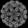 40049MC M: map data used to model this data C: citing same article ( |
|---|---|
| Similar structure data |

-Links
 PDBj
PDBj

- Assembly
| Deposited unit | 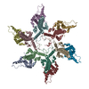
|
|---|---|
| 1 |
|
-Components
| #1: Protein | Mass: 16840.186 Da / Num. of mol.: 12 Source method: isolated from a genetically manipulated source Source: (gene. exp.) Hepatitis B virus / Production host:  |
|---|
-Experimental details
-Experiment
| Experiment | Method: ELECTRON MICROSCOPY |
|---|---|
| EM experiment | Aggregation state: PARTICLE / 3D reconstruction method: single particle reconstruction |
- Sample preparation
| Component | Name: Reassembled HBV empty Cp183 capsid with importin-beta / Type: COMPLEX / Details: Core protein Cp183 expressed in E coli / Entity ID: all / Source: RECOMBINANT |
|---|---|
| Molecular weight | Experimental value: NO |
| Source (natural) | Organism: Hepatitis B virus |
| Source (recombinant) | Organism: |
| Details of virus | Empty: NO / Enveloped: NO / Isolate: OTHER / Type: VIRUS-LIKE PARTICLE |
| Buffer solution | pH: 7.5 / Details: 0.15 M NaCl, 20 mM Tris-HCl pH 7.5, and 10 mM DTT |
| Specimen | Embedding applied: NO / Shadowing applied: NO / Staining applied: NO / Vitrification applied: YES / Details: Empty HBV capsid : Importin-beta sample at 1:205 |
| Vitrification | Instrument: FEI VITROBOT MARK IV / Cryogen name: ETHANE / Humidity: 100 % |
- Electron microscopy imaging
| Experimental equipment |  Model: Talos Arctica / Image courtesy: FEI Company |
|---|---|
| Microscopy | Model: FEI TALOS ARCTICA |
| Electron gun | Electron source:  FIELD EMISSION GUN / Accelerating voltage: 200 kV / Illumination mode: FLOOD BEAM FIELD EMISSION GUN / Accelerating voltage: 200 kV / Illumination mode: FLOOD BEAM |
| Electron lens | Mode: BRIGHT FIELD / Nominal defocus max: 4000 nm / Nominal defocus min: 800 nm |
| Image recording | Electron dose: 30 e/Å2 / Detector mode: COUNTING / Film or detector model: FEI FALCON III (4k x 4k) |
- Processing
| Software | Name: PHENIX / Version: 1.20.1_4487: / Classification: refinement | |||||||||||||||||||||||||||||||||
|---|---|---|---|---|---|---|---|---|---|---|---|---|---|---|---|---|---|---|---|---|---|---|---|---|---|---|---|---|---|---|---|---|---|---|
| EM software |
| |||||||||||||||||||||||||||||||||
| CTF correction | Type: PHASE FLIPPING ONLY | |||||||||||||||||||||||||||||||||
| Particle selection | Num. of particles selected: 31166 | |||||||||||||||||||||||||||||||||
| Symmetry | Point symmetry: C1 (asymmetric) | |||||||||||||||||||||||||||||||||
| 3D reconstruction | Resolution: 4 Å / Resolution method: FSC 0.143 CUT-OFF / Num. of particles: 1154892 / Symmetry type: POINT | |||||||||||||||||||||||||||||||||
| Atomic model building | Protocol: RIGID BODY FIT Details: A hexamer model was flexibly fitted into the hexamer maps using Alphafold, ISOLDE, PHENIX, and Coot. | |||||||||||||||||||||||||||||||||
| Refine LS restraints |
|
