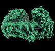
 Movie
Movie Controller

[English] 日本語
 Yorodumi
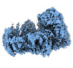
- PDB-8g5o: Cryo-EM structure of the Guide loop Engagement Complex (IV) of Hu... -
+ Open data

- Basic information
| Entry | Database: PDB / ID: 8g5o | ||||||
|---|---|---|---|---|---|---|---|
| Title | Cryo-EM structure of the Guide loop Engagement Complex (IV) of Human Mitochondrial DNA Polymerase Gamma | ||||||
 Components Components |
| ||||||
 Keywords Keywords | REPLICATION/DNA/RNA / Mitochondrial DNA Polymerase / DNA Proofreading / Guide loop Engagement / REPLICATION-DNA-RNA complex | ||||||
| Function / homology |  Function and homology information Function and homology informationgamma DNA polymerase complex / mitochondrial DNA replication / positive regulation of DNA-directed DNA polymerase activity / single-stranded DNA 3'-5' DNA exonuclease activity / Hydrolases; Acting on ester bonds; Exodeoxyribonucleases producing 5'-phosphomonoesters / DNA replication proofreading / DNA metabolic process / DNA polymerase processivity factor activity / mitochondrial nucleoid / Lyases; Carbon-oxygen lyases; Other carbon-oxygen lyases ...gamma DNA polymerase complex / mitochondrial DNA replication / positive regulation of DNA-directed DNA polymerase activity / single-stranded DNA 3'-5' DNA exonuclease activity / Hydrolases; Acting on ester bonds; Exodeoxyribonucleases producing 5'-phosphomonoesters / DNA replication proofreading / DNA metabolic process / DNA polymerase processivity factor activity / mitochondrial nucleoid / Lyases; Carbon-oxygen lyases; Other carbon-oxygen lyases / 5'-deoxyribose-5-phosphate lyase activity / DNA polymerase binding / 3'-5' exonuclease activity / base-excision repair, gap-filling / Transcriptional activation of mitochondrial biogenesis / base-excision repair / DNA-templated DNA replication / double-stranded DNA binding / protease binding / in utero embryonic development / DNA-directed DNA polymerase / DNA-directed DNA polymerase activity / mitochondrial matrix / intracellular membrane-bounded organelle / chromatin binding / protein-containing complex / mitochondrion / DNA binding / identical protein binding / cytoplasm Similarity search - Function | ||||||
| Biological species |  Homo sapiens (human) Homo sapiens (human)synthetic RNA (others) synthetic construct (others) | ||||||
| Method | ELECTRON MICROSCOPY / single particle reconstruction / cryo EM / Resolution: 2.61 Å | ||||||
 Authors Authors | Nayak, A.R. / Buchel, G. / Herbine, K.H. / Sarfallah, A. / Sokolova, V.O. / Zamudio-Ochoa, A. / Temiakov, D. | ||||||
| Funding support |  United States, 1items United States, 1items
| ||||||
 Citation Citation | Journal: Nat Commun / Year: 2023 Title: Structural basis for DNA proofreading. Authors: Gina Buchel / Ashok R Nayak / Karl Herbine / Azadeh Sarfallah / Viktoriia O Sokolova / Angelica Zamudio-Ochoa / Dmitry Temiakov / Abstract: DNA polymerase (DNAP) can correct errors in DNA during replication by proofreading, a process critical for cell viability. However, the mechanism by which an erroneously incorporated base ...DNA polymerase (DNAP) can correct errors in DNA during replication by proofreading, a process critical for cell viability. However, the mechanism by which an erroneously incorporated base translocates from the polymerase to the exonuclease site and the corrected DNA terminus returns has remained elusive. Here, we present an ensemble of nine high-resolution structures representing human mitochondrial DNA polymerase Gamma, Polγ, captured during consecutive proofreading steps. The structures reveal key events, including mismatched base recognition, its dissociation from the polymerase site, forward translocation of DNAP, alterations in DNA trajectory, repositioning and refolding of elements for primer separation, DNAP backtracking, and displacement of the mismatched base into the exonuclease site. Altogether, our findings suggest a conserved 'bolt-action' mechanism of proofreading based on iterative cycles of DNAP translocation without dissociation from the DNA, facilitating primer transfer between catalytic sites. Functional assays and mutagenesis corroborate this mechanism, connecting pathogenic mutations to crucial structural elements in proofreading steps. | ||||||
| History |
|
- Structure visualization
| Structure viewer | Molecule: MolmilJmol/JSmol |
|---|
- Downloads & links
-Download
| PDBx/mmCIF format | 8g5o.cif.gz | 341.4 KB | Display | PDBx/mmCIF format |
|---|---|---|---|---|
| PDB format | pdb8g5o.ent.gz | 246.7 KB | Display | PDB format |
| PDBx/mmJSON format | 8g5o.json.gz | Tree view | PDBx/mmJSON format | |
| Others |  Other downloads Other downloads |
-Validation report
| Summary document | 8g5o_validation.pdf.gz | 1.5 MB | Display | wwPDB validaton report |
|---|---|---|---|---|
| Full document | 8g5o_full_validation.pdf.gz | 1.6 MB | Display | |
| Data in XML | 8g5o_validation.xml.gz | 57.5 KB | Display | |
| Data in CIF | 8g5o_validation.cif.gz | 89 KB | Display | |
| Arichive directory | https://data.pdbj.org/pub/pdb/validation_reports/g5/8g5oftp://data.pdbj.org/pub/pdb/validation_reports/g5/8g5o | HTTPS FTP |
-Related structure data
| Related structure data |  29751MC 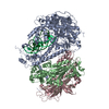 8g5iC 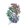 8g5jC 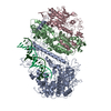 8g5kC 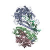 8g5lC 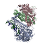 8g5mC 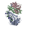 8g5nC 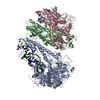 8g5pC 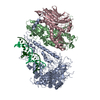 8t7eC M: map data used to model this data C: citing same article ( |
|---|---|
| Similar structure data |
-Links
 PDBj
PDBj


- Assembly
| Deposited unit | 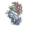
|
|---|---|
| 1 |
|
-Components
| #1: Protein | Mass: 139628.672 Da / Num. of mol.: 1 / Mutation: D198A, E200A Source method: isolated from a genetically manipulated source Source: (gene. exp.) Homo sapiens (human) / Gene: POLG, MDP1, POLG1, POLGA / Plasmid: pFastBac1 / Cell line (production host): Sf9 / Production host:   Spodoptera frugiperda (fall armyworm) / Tissue (production host): Ovary / References: UniProt: P54098, DNA-directed DNA polymerase Spodoptera frugiperda (fall armyworm) / Tissue (production host): Ovary / References: UniProt: P54098, DNA-directed DNA polymerase | ||||
|---|---|---|---|---|---|
| #2: Protein | Mass: 54991.000 Da / Num. of mol.: 2 Source method: isolated from a genetically manipulated source Source: (gene. exp.) Homo sapiens (human) / Gene: POLG2, MTPOLB / Plasmid: pProEX / Production host:  #3: RNA chain | | Mass: 7857.758 Da / Num. of mol.: 1 / Source method: obtained synthetically / Source: (synth.) synthetic RNA (others) #4: DNA chain | | Mass: 9176.879 Da / Num. of mol.: 1 / Source method: obtained synthetically / Source: (synth.) synthetic construct (others) |
-Experimental details
-Experiment
| Experiment | Method: ELECTRON MICROSCOPY |
|---|---|
| EM experiment | Aggregation state: PARTICLE / 3D reconstruction method: single particle reconstruction |
- Sample preparation
| Component | Name: Cryo-EM structure of the Guide Loop Engagement Complex (IV) of Human Mitochondrial DNA Polymerase Gamma Type: COMPLEX Details: Human mitochondrial DNA polymerase PolG (exonuclease deficient D198A/E200A variant) assembled on an RNA-DNA scaffold in the presence of GTP Entity ID: all / Source: RECOMBINANT |
|---|---|
| Molecular weight | Value: 0.362 MDa / Experimental value: YES |
| Source (natural) | Organism: Homo sapiens (human) |
| Source (recombinant) | Organism: Spodoptera frugiperda (fall armyworm) |
| Buffer solution | pH: 7.9 Details: 10 mM Tris-Hcl pH 7.9, 100 mM Nacl, 10 mM DTT, and 2 mM MgCl2 |
| Specimen | Conc.: 0.52 mg/ml / Embedding applied: NO / Shadowing applied: NO / Staining applied: NO / Vitrification applied: YES Details: 1:1 complex of Exo- PolG and RNA-DNA scaffold in the presence of 0.1 mM dGTP |
| Specimen support | Grid material: GOLD / Grid type: UltrAuFoil R1.2/1.3 |
| Vitrification | Instrument: FEI VITROBOT MARK IV / Cryogen name: ETHANE / Humidity: 100 % / Chamber temperature: 277 K |
- Electron microscopy imaging
| Experimental equipment |  Model: Titan Krios / Image courtesy: FEI Company |
|---|---|
| Microscopy | Model: FEI TITAN KRIOS |
| Electron gun | Electron source:  FIELD EMISSION GUN / Accelerating voltage: 300 kV / Illumination mode: FLOOD BEAM FIELD EMISSION GUN / Accelerating voltage: 300 kV / Illumination mode: FLOOD BEAM |
| Electron lens | Mode: BRIGHT FIELD / Nominal magnification: 105000 X / Nominal defocus max: 2000 nm / Nominal defocus min: 800 nm / Cs: 2.7 mm |
| Specimen holder | Cryogen: NITROGEN |
| Image recording | Electron dose: 60 e/Å2 / Film or detector model: GATAN K3 (6k x 4k) / Num. of grids imaged: 2 / Num. of real images: 12937 |
| EM imaging optics | Energyfilter slit width: 20 eV |
- Processing
| EM software |
| ||||||||||||||||||||||||||||||||
|---|---|---|---|---|---|---|---|---|---|---|---|---|---|---|---|---|---|---|---|---|---|---|---|---|---|---|---|---|---|---|---|---|---|
| CTF correction | Type: PHASE FLIPPING AND AMPLITUDE CORRECTION | ||||||||||||||||||||||||||||||||
| Particle selection | Num. of particles selected: 14667792 | ||||||||||||||||||||||||||||||||
| Symmetry | Point symmetry: C1 (asymmetric) | ||||||||||||||||||||||||||||||||
| 3D reconstruction | Resolution: 2.61 Å / Resolution method: FSC 0.143 CUT-OFF / Num. of particles: 426672 / Num. of class averages: 1 / Symmetry type: POINT | ||||||||||||||||||||||||||||||||
| Atomic model building | Protocol: FLEXIBLE FIT / Space: REAL | ||||||||||||||||||||||||||||||||
| Refine LS restraints |
|
