 Movie
Movie Controller


+ Open data

- Basic information
| Entry | Database: PDB / ID: 8fzn | |||||||||
|---|---|---|---|---|---|---|---|---|---|---|
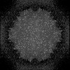
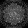
| Title | Cryo-EM Structure of AAV2-R404A Variant | |||||||||
 Components Components | Capsid protein VP1 | |||||||||
 Keywords Keywords | VIRUS / Symmetry / capsid / AAV2 | |||||||||
| Function / homology |  Function and homology information Function and homology informationsymbiont entry into host cell via permeabilization of host membrane / host cell nucleolus / T=1 icosahedral viral capsid / clathrin-dependent endocytosis of virus by host cell / virion attachment to host cell / structural molecule activity Similarity search - Function | |||||||||
| Biological species |   Adeno-associated virus Adeno-associated virus | |||||||||
| Method | ELECTRON MICROSCOPY / single particle reconstruction / cryo EM / Resolution: 3.62 Å | |||||||||
 Authors Authors | Bennett, A.D. / McKenna, R. | |||||||||
| Funding support |  United States, 2items United States, 2items
| |||||||||
 Citation Citation | Journal: to be published Title: Cryo-EM Structure of genome containing AAV2 Authors: Bennett, A.D. / Mckenna, R. | |||||||||
| History |
|
- Structure visualization
| Structure viewer | Molecule: MolmilJmol/JSmol |
|---|
- Downloads & links
-Download
| PDBx/mmCIF format | 8fzn.cif.gz | 5.5 MB | Display | PDBx/mmCIF format |
|---|---|---|---|---|
| PDB format | pdb8fzn.ent.gz | Display | PDB format | |
| PDBx/mmJSON format | 8fzn.json.gz | Tree view | PDBx/mmJSON format | |
| Others |  Other downloads Other downloads |
-Validation report
| Arichive directory | https://data.pdbj.org/pub/pdb/validation_reports/fz/8fznftp://data.pdbj.org/pub/pdb/validation_reports/fz/8fzn | HTTPS FTP |
|---|
-Related structure data
| Related structure data | 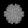 29636MC  8fywC  8fz0C M: map data used to model this data C: citing same article ( |
|---|---|
| Similar structure data |
-Links
 PDBj
PDBj


- Assembly
| Deposited unit | 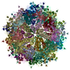
|
|---|---|
| 1 |
|
-Components
| #1: Protein | Mass: 56722.445 Da / Num. of mol.: 60 Source method: isolated from a genetically manipulated source Source: (gene. exp.) Adeno-associated virus / Gene: VP1 / Variant: AAV2-R404A / Production host:   Spodoptera frugiperda (fall armyworm) / References: UniProt: P03135 Spodoptera frugiperda (fall armyworm) / References: UniProt: P03135 |
|---|
-Experimental details
-Experiment
| Experiment | Method: ELECTRON MICROSCOPY |
|---|---|
| EM experiment | Aggregation state: PARTICLE / 3D reconstruction method: single particle reconstruction |
- Sample preparation
| Component | Name: adeno-associated virus 2 / Type: VIRUS / Entity ID: all / Source: RECOMBINANT |
|---|---|
| Molecular weight | Value: 3.9 MDa / Experimental value: NO |
| Source (natural) | Organism: adeno-associated virus 2 |
| Source (recombinant) | Organism: Baculovirus expression vector pCTdual |
| Details of virus | Empty: NO / Enveloped: NO / Isolate: SEROTYPE / Type: VIRION |
| Natural host | Organism: Homo sapiens |
| Virus shell | Name: capsid / Diameter: 26 nm / Triangulation number (T number): 1 |
| Buffer solution | pH: 7.3 / Details: 1XPBS, 1 mM MgCl2, 2.5 mM KCl pH7.3 |
| Buffer component | Conc.: 1 mM / Name: TD / Formula: PBS-MK |
| Specimen | Conc.: 0.5 mg/ml / Embedding applied: NO / Shadowing applied: NO / Staining applied: NO / Vitrification applied: YES |
| Specimen support | Grid type: Quantifoil |
| Vitrification | Instrument: FEI VITROBOT MARK IV / Cryogen name: ETHANE |
- Electron microscopy imaging
| Experimental equipment |  Model: Titan Krios / Image courtesy: FEI Company |
|---|---|
| Microscopy | Model: FEI TITAN KRIOS |
| Electron gun | Electron source:  FIELD EMISSION GUN / Accelerating voltage: 300 kV / Illumination mode: SPOT SCAN FIELD EMISSION GUN / Accelerating voltage: 300 kV / Illumination mode: SPOT SCAN |
| Electron lens | Mode: BRIGHT FIELD / Nominal defocus max: 2500 nm / Nominal defocus min: 1000 nm / Cs: 2.7 mm |
| Image recording | Electron dose: 60 e/Å2 / Film or detector model: DIRECT ELECTRON DE-64 (8k x 8k) / Num. of real images: 1674 |
- Processing
| Software | Name: PHENIX / Version: 1.10-2155_2155: / Classification: refinement | ||||||||||||||||||||||||
|---|---|---|---|---|---|---|---|---|---|---|---|---|---|---|---|---|---|---|---|---|---|---|---|---|---|
| EM software |
| ||||||||||||||||||||||||
| CTF correction | Type: PHASE FLIPPING AND AMPLITUDE CORRECTION | ||||||||||||||||||||||||
| 3D reconstruction | Resolution: 3.62 Å / Resolution method: FSC 0.143 CUT-OFF / Num. of particles: 23783 / Symmetry type: POINT | ||||||||||||||||||||||||
| Atomic model building | Protocol: AB INITIO MODEL | ||||||||||||||||||||||||
| Refine LS restraints |
|
