Movie
Movie Controller


+ Open data

- Basic information
| Entry | Database: PDB / ID: 8fzc | ||||||||||||||||||||||||
|---|---|---|---|---|---|---|---|---|---|---|---|---|---|---|---|---|---|---|---|---|---|---|---|---|---|
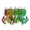
| Title | HIV-2 Gag Capsid from Immature Virus-like Particles | ||||||||||||||||||||||||
 Components Components | Spacer peptide 2 | ||||||||||||||||||||||||
 Keywords Keywords | VIRUS LIKE PARTICLE / Retrovirus / Lentivirus / Gag / Capsid / HIV-2 / lattice | ||||||||||||||||||||||||
| Function / homology |  Function and homology information Function and homology informationviral budding via host ESCRT complex / host multivesicular body / viral nucleocapsid / viral translational frameshifting / host cell plasma membrane / host cell nucleus / virion membrane / structural molecule activity / RNA binding / zinc ion binding Similarity search - Function | ||||||||||||||||||||||||
| Biological species |  Human immunodeficiency virus type 2 Human immunodeficiency virus type 2 | ||||||||||||||||||||||||
| Method | ELECTRON MICROSCOPY / single particle reconstruction / cryo EM / Resolution: 5.5 Å | ||||||||||||||||||||||||
 Authors Authors | Talledge, N. / Zhang, W. / Mansky, L.M. | ||||||||||||||||||||||||
| Funding support |  United States, 7items United States, 7items
| ||||||||||||||||||||||||
 Citation Citation | Journal: J Mol Biol / Year: 2023 Title: HIV-2 Immature Particle Morphology Provides Insights into Gag Lattice Stability and Virus Maturation. Authors: Nathaniel Talledge / Huixin Yang / Ke Shi / Raffaele Coray / Guichuan Yu / William G Arndt / Shuyu Meng / Gloria C Baxter / Luiza M Mendonça / Daniel Castaño-Díez / Hideki Aihara / Louis ...Authors: Nathaniel Talledge / Huixin Yang / Ke Shi / Raffaele Coray / Guichuan Yu / William G Arndt / Shuyu Meng / Gloria C Baxter / Luiza M Mendonça / Daniel Castaño-Díez / Hideki Aihara / Louis M Mansky / Wei Zhang /   Abstract: Retrovirus immature particle morphology consists of a membrane enclosed, pleomorphic, spherical and incomplete lattice of Gag hexamers. Previously, we demonstrated that human immunodeficiency virus ...Retrovirus immature particle morphology consists of a membrane enclosed, pleomorphic, spherical and incomplete lattice of Gag hexamers. Previously, we demonstrated that human immunodeficiency virus type 2 (HIV-2) immature particles possess a distinct and extensive Gag lattice morphology. To better understand the nature of the continuously curved hexagonal Gag lattice, we have used the single particle cryo-electron microscopy method to determine the HIV-2 Gag lattice structure for immature virions. The reconstruction map at 5.5 Å resolution revealed a stable, wineglass-shaped Gag hexamer structure with structural features consistent with other lentiviral immature Gag lattice structures. Cryo-electron tomography provided evidence for nearly complete ordered Gag lattice structures in HIV-2 immature particles. We also solved a 1.98 Å resolution crystal structure of the carboxyl-terminal domain (CTD) of the HIV-2 capsid (CA) protein that identified a structured helix 12 supported via an interaction of helix 10 in the absence of the SP1 region of Gag. Residues at the helix 10-12 interface proved critical in maintaining HIV-2 particle release and infectivity. Taken together, our findings provide the first 3D organization of HIV-2 immature Gag lattice and important insights into both HIV Gag lattice stabilization and virus maturation. | ||||||||||||||||||||||||
| History |
|
- Structure visualization
| Structure viewer | Molecule: MolmilJmol/JSmol |
|---|
- Downloads & links
-Download
| PDBx/mmCIF format | 8fzc.cif.gz | 124.5 KB | Display | PDBx/mmCIF format |
|---|---|---|---|---|
| PDB format | pdb8fzc.ent.gz | 98.3 KB | Display | PDB format |
| PDBx/mmJSON format | 8fzc.json.gz | Tree view | PDBx/mmJSON format | |
| Others |  Other downloads Other downloads |
-Validation report
| Arichive directory | https://data.pdbj.org/pub/pdb/validation_reports/fz/8fzcftp://data.pdbj.org/pub/pdb/validation_reports/fz/8fzc | HTTPS FTP |
|---|
-Related structure data
| Related structure data | 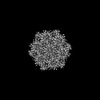 29607MC  7tv2C C: citing same article ( M: map data used to model this data |
|---|---|
| Similar structure data |
-Links
 PDBj
PDBj
- Assembly
| Deposited unit | 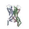
|
|---|---|
| 1 | x 6
|
-Components
| #1: Protein | Mass: 25109.822 Da / Num. of mol.: 3 Source method: isolated from a genetically manipulated source Source: (gene. exp.) Human immunodeficiency virus type 2 (ISOLATE ROD)Gene: gag / Variant: isolate ROD / Plasmid: pN3 / Details (production host): CMV promotor / Cell line (production host): HEK293 / Production host:  Homo sapiens (human) / References: UniProt: P04590 Homo sapiens (human) / References: UniProt: P04590 |
|---|
-Experimental details
-Experiment
| Experiment | Method: ELECTRON MICROSCOPY |
|---|---|
| EM experiment | Aggregation state: PARTICLE / 3D reconstruction method: single particle reconstruction |
- Sample preparation
| Component | Name: Human immunodeficiency virus type 2 (ISOLATE ROD) / Type: VIRUS Details: HIV-2 VLPs generated by Gag co-overexpressed with HIV-2 Env in Hek293T cells and purified from media supernatant Entity ID: all / Source: RECOMBINANT | ||||||||||||||||||||
|---|---|---|---|---|---|---|---|---|---|---|---|---|---|---|---|---|---|---|---|---|---|
| Molecular weight | Experimental value: NO | ||||||||||||||||||||
| Source (natural) | Organism: Human immunodeficiency virus type 2 (ISOLATE ROD) | ||||||||||||||||||||
| Source (recombinant) | Organism: Homo sapiens (human) | ||||||||||||||||||||
| Details of virus | Empty: NO / Enveloped: YES / Isolate: STRAIN / Type: VIRUS-LIKE PARTICLE | ||||||||||||||||||||
| Natural host | Organism: Homo sapiens | ||||||||||||||||||||
| Virus shell | Name: Capsid / Diameter: 2100 nm | ||||||||||||||||||||
| Buffer solution | pH: 8 / Details: 100 mM NaCl, 10 mM Tris-HCL pH 8.0, 1mM EDTA | ||||||||||||||||||||
| Buffer component |
| ||||||||||||||||||||
| Specimen | Conc.: 10 mg/ml / Embedding applied: NO / Shadowing applied: NO / Staining applied: NO / Vitrification applied: YES | ||||||||||||||||||||
| Specimen support | Details: Leica ACE600 Glow Discharger. / Grid material: COPPER / Grid mesh size: 200 divisions/in. / Grid type: Quantifoil R2/2 | ||||||||||||||||||||
| Vitrification | Instrument: LEICA EM GP / Cryogen name: ETHANE / Humidity: 85 % / Chamber temperature: 292.15 K / Details: Leica GP2 Grid Plunger. |
- Electron microscopy imaging
| Experimental equipment |  Model: Titan Krios / Image courtesy: FEI Company |
|---|---|
| Microscopy | Model: FEI TITAN KRIOS |
| Electron gun | Electron source:  FIELD EMISSION GUN / Accelerating voltage: 300 kV / Illumination mode: FLOOD BEAM FIELD EMISSION GUN / Accelerating voltage: 300 kV / Illumination mode: FLOOD BEAM |
| Electron lens | Mode: BRIGHT FIELD / Nominal magnification: 105000 X / Nominal defocus max: 2250 nm / Nominal defocus min: 750 nm / Alignment procedure: COMA FREE |
| Specimen holder | Cryogen: NITROGEN / Specimen holder model: FEI TITAN KRIOS AUTOGRID HOLDER |
| Image recording | Average exposure time: 10 sec. / Electron dose: 50 e/Å2 / Detector mode: SUPER-RESOLUTION / Film or detector model: GATAN K2 SUMMIT (4k x 4k) / Num. of grids imaged: 10 / Num. of real images: 2508 |
| EM imaging optics | Energyfilter slit width: 20 eV |
| Image scans | Movie frames/image: 40 / Used frames/image: 1-40 |
- Processing
| EM software |
| ||||||||||||||||||||||||||||||||||||||||
|---|---|---|---|---|---|---|---|---|---|---|---|---|---|---|---|---|---|---|---|---|---|---|---|---|---|---|---|---|---|---|---|---|---|---|---|---|---|---|---|---|---|
| CTF correction | Type: PHASE FLIPPING AND AMPLITUDE CORRECTION | ||||||||||||||||||||||||||||||||||||||||
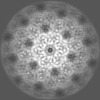
| Symmetry | Point symmetry: C6 (6 fold cyclic) | ||||||||||||||||||||||||||||||||||||||||
| 3D reconstruction | Resolution: 5.5 Å / Resolution method: FSC 0.143 CUT-OFF / Num. of particles: 46017 / Algorithm: FOURIER SPACE / Num. of class averages: 1 / Symmetry type: POINT | ||||||||||||||||||||||||||||||||||||||||
| Atomic model building | Protocol: RIGID BODY FIT | ||||||||||||||||||||||||||||||||||||||||
| Atomic model building | 3D fitting-ID: 1 / Chain-ID: A / Pdb chain-ID: A / Source name: PDB / Type: experimental model
| ||||||||||||||||||||||||||||||||||||||||
| Refine LS restraints |
|