[English] 日本語
 Yorodumi
- PDB-8fop: Structure of Agrobacterium tumefaciens bacteriophage Milano curve... -
+ Open data
- Basic information
| Entry | Database: PDB / ID: 8fop | ||||||
|---|---|---|---|---|---|---|---|
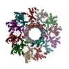
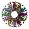
| Title | Structure of Agrobacterium tumefaciens bacteriophage Milano curved tail | ||||||
 Components Components |
| ||||||
 Keywords Keywords | VIRUS / Myophage / redox trigger | ||||||
| Function / homology | Tail sheath protein / Virion-associated protein Function and homology information Function and homology information | ||||||
| Biological species |  Agrobacterium phage Milano (virus) Agrobacterium phage Milano (virus) | ||||||
| Method | ELECTRON MICROSCOPY / single particle reconstruction / cryo EM / Resolution: 3.2 Å | ||||||
 Authors Authors | Sonani, R.R. / Leiman, P.G. / Wang, F. / Kreutzberger, M.A.B. / Sebastian, A. / Esteves, N.C. / Kelly, R.J. / Scharf, B. / Egelman, E.H. | ||||||
| Funding support |  United States, 1items United States, 1items
| ||||||
 Citation Citation |  Journal: Nat Commun / Year: 2024 Journal: Nat Commun / Year: 2024Title: An extensive disulfide bond network prevents tail contraction in Agrobacterium tumefaciens phage Milano. Authors: Ravi R Sonani / Lee K Palmer / Nathaniel C Esteves / Abigail A Horton / Amanda L Sebastian / Rebecca J Kelly / Fengbin Wang / Mark A B Kreutzberger / William K Russell / Petr G Leiman / ...Authors: Ravi R Sonani / Lee K Palmer / Nathaniel C Esteves / Abigail A Horton / Amanda L Sebastian / Rebecca J Kelly / Fengbin Wang / Mark A B Kreutzberger / William K Russell / Petr G Leiman / Birgit E Scharf / Edward H Egelman /  Abstract: A contractile sheath and rigid tube assembly is a widespread apparatus used by bacteriophages, tailocins, and the bacterial type VI secretion system to penetrate cell membranes. In this mechanism, ...A contractile sheath and rigid tube assembly is a widespread apparatus used by bacteriophages, tailocins, and the bacterial type VI secretion system to penetrate cell membranes. In this mechanism, contraction of an external sheath powers the motion of an inner tube through the membrane. The structure, energetics, and mechanism of the machinery imply rigidity and straightness. The contractile tail of Agrobacterium tumefaciens bacteriophage Milano is flexible and bent to varying degrees, which sets it apart from other contractile tail-like systems. Here, we report structures of the Milano tail including the sheath-tube complex, baseplate, and putative receptor-binding proteins. The flexible-to-rigid transformation of the Milano tail upon contraction can be explained by unique electrostatic properties of the tail tube and sheath. All components of the Milano tail, including sheath subunits, are crosslinked by disulfides, some of which must be reduced for contraction to occur. The putative receptor-binding complex of Milano contains a tailspike, a tail fiber, and at least two small proteins that form a garland around the distal ends of the tailspikes and tail fibers. Despite being flagellotropic, Milano lacks thread-like tail filaments that can wrap around the flagellum, and is thus likely to employ a different binding mechanism. | ||||||
| History |
|
- Structure visualization
| Structure viewer | Molecule:  Molmil Molmil Jmol/JSmol Jmol/JSmol |
|---|
- Downloads & links
- Download
| PDBx/mmCIF format |  8fop.cif.gz 8fop.cif.gz | 1.2 MB | Display |  PDBx/mmCIF format PDBx/mmCIF format |
|---|---|---|---|---|
| PDB format |  pdb8fop.ent.gz pdb8fop.ent.gz | 992.8 KB | Display |  PDB format PDB format |
| PDBx/mmJSON format |  8fop.json.gz 8fop.json.gz | Tree view |  PDBx/mmJSON format PDBx/mmJSON format | |
| Others |  Other downloads Other downloads |
-Validation report
| Summary document |  8fop_validation.pdf.gz 8fop_validation.pdf.gz | 1.8 MB | Display |  wwPDB validaton report wwPDB validaton report |
|---|---|---|---|---|
| Full document |  8fop_full_validation.pdf.gz 8fop_full_validation.pdf.gz | 1.9 MB | Display | |
| Data in XML |  8fop_validation.xml.gz 8fop_validation.xml.gz | 179.9 KB | Display | |
| Data in CIF |  8fop_validation.cif.gz 8fop_validation.cif.gz | 282.8 KB | Display | |
| Arichive directory |  https://data.pdbj.org/pub/pdb/validation_reports/fo/8fop https://data.pdbj.org/pub/pdb/validation_reports/fo/8fop ftp://data.pdbj.org/pub/pdb/validation_reports/fo/8fop ftp://data.pdbj.org/pub/pdb/validation_reports/fo/8fop | HTTPS FTP |
-Related structure data
| Related structure data |  29353MC  8fouC  8foyC  8fqcC M: map data used to model this data C: citing same article ( |
|---|---|
| Similar structure data | Similarity search - Function & homology  F&H Search F&H Search |
- Links
Links
- Assembly
| Deposited unit | 
|
|---|---|
| 1 |
|
- Components
| #1: Protein | Mass: 14673.427 Da / Num. of mol.: 18 / Source method: isolated from a natural source / Source: (natural)  Agrobacterium phage Milano (virus) / References: UniProt: A0A482MHE7 Agrobacterium phage Milano (virus) / References: UniProt: A0A482MHE7#2: Protein | Mass: 53896.094 Da / Num. of mol.: 12 / Source method: isolated from a natural source / Source: (natural)  Agrobacterium phage Milano (virus) / References: UniProt: A0A482MFS8 Agrobacterium phage Milano (virus) / References: UniProt: A0A482MFS8Has protein modification | Y | |
|---|
-Experimental details
-Experiment
| Experiment | Method: ELECTRON MICROSCOPY |
|---|---|
| EM experiment | Aggregation state: PARTICLE / 3D reconstruction method: single particle reconstruction |
- Sample preparation
| Component | Name: Agrobacterium phage Milano / Type: VIRUS / Entity ID: all / Source: NATURAL |
|---|---|
| Source (natural) | Organism:  Agrobacterium phage Milano (virus) Agrobacterium phage Milano (virus) |
| Details of virus | Empty: NO / Enveloped: YES / Isolate: SPECIES / Type: VIRION |
| Natural host | Organism: Agrobacterium fabrum str. C58 |
| Buffer solution | pH: 7.4 |
| Specimen | Embedding applied: NO / Shadowing applied: NO / Staining applied: NO / Vitrification applied: YES |
| Vitrification | Cryogen name: ETHANE |
- Electron microscopy imaging
| Experimental equipment |  Model: Titan Krios / Image courtesy: FEI Company |
|---|---|
| Microscopy | Model: TFS KRIOS |
| Electron gun | Electron source:  FIELD EMISSION GUN / Accelerating voltage: 300 kV / Illumination mode: FLOOD BEAM FIELD EMISSION GUN / Accelerating voltage: 300 kV / Illumination mode: FLOOD BEAM |
| Electron lens | Mode: BRIGHT FIELD / Nominal defocus max: 2200 nm / Nominal defocus min: 1200 nm |
| Image recording | Electron dose: 50 e/Å2 / Film or detector model: GATAN K3 (6k x 4k) |
- Processing
| CTF correction | Type: PHASE FLIPPING AND AMPLITUDE CORRECTION | ||||||||||||||||||||||||
|---|---|---|---|---|---|---|---|---|---|---|---|---|---|---|---|---|---|---|---|---|---|---|---|---|---|
| 3D reconstruction | Resolution: 3.2 Å / Resolution method: FSC 0.143 CUT-OFF / Num. of particles: 228393 / Symmetry type: POINT | ||||||||||||||||||||||||
| Refine LS restraints |
|
 Movie
Movie Controller


 PDBj
PDBj