 Movie
Movie Controller


+ Open data

- Basic information
| Entry | Database: PDB / ID: 8fbb | ||||||||||||||||||
|---|---|---|---|---|---|---|---|---|---|---|---|---|---|---|---|---|---|---|---|
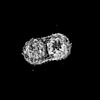
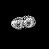
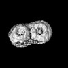
| Title | LH2-LH3 antenna in parallel configuration embedded in a nanodisc | ||||||||||||||||||
 Components Components |
| ||||||||||||||||||
 Keywords Keywords | PHOTOSYNTHESIS / antenna / membrane protein / nanodisc / bacteriochlorophyll | ||||||||||||||||||
| Function / homology |  Function and homology information Function and homology informationorganelle inner membrane / plasma membrane light-harvesting complex / bacteriochlorophyll binding / photosynthesis, light reaction / metal ion binding / plasma membrane Similarity search - Function | ||||||||||||||||||
| Biological species |  Magnetospirillum molischianum (magnetotactic) Magnetospirillum molischianum (magnetotactic) | ||||||||||||||||||
| Method | ELECTRON MICROSCOPY / single particle reconstruction / cryo EM / Resolution: 11.3 Å | ||||||||||||||||||
 Authors Authors | Toporik, H. / Harris, D. / Schlau-Cohen, G.S. / Mazor, Y. | ||||||||||||||||||
| Funding support |  United States, 5items United States, 5items
| ||||||||||||||||||
 Citation Citation | Journal: Proc Natl Acad Sci U S A / Year: 2023 Title: Elucidating interprotein energy transfer dynamics within the antenna network from purple bacteria. Authors: Dihao Wang / Olivia C Fiebig / Dvir Harris / Hila Toporik / Yi Ji / Chern Chuang / Muath Nairat / Ashley L Tong / John I Ogren / Stephanie M Hart / Jianshu Cao / James N Sturgis / Yuval ...Authors: Dihao Wang / Olivia C Fiebig / Dvir Harris / Hila Toporik / Yi Ji / Chern Chuang / Muath Nairat / Ashley L Tong / John I Ogren / Stephanie M Hart / Jianshu Cao / James N Sturgis / Yuval Mazor / Gabriela S Schlau-Cohen /  Abstract: In photosynthesis, absorbed light energy transfers through a network of antenna proteins with near-unity quantum efficiency to reach the reaction center, which initiates the downstream biochemical ...In photosynthesis, absorbed light energy transfers through a network of antenna proteins with near-unity quantum efficiency to reach the reaction center, which initiates the downstream biochemical reactions. While the energy transfer dynamics within individual antenna proteins have been extensively studied over the past decades, the dynamics between the proteins are poorly understood due to the heterogeneous organization of the network. Previously reported timescales averaged over such heterogeneity, obscuring individual interprotein energy transfer steps. Here, we isolated and interrogated interprotein energy transfer by embedding two variants of the primary antenna protein from purple bacteria, light-harvesting complex 2 (LH2), together into a near-native membrane disc, known as a nanodisc. We integrated ultrafast transient absorption spectroscopy, quantum dynamics simulations, and cryogenic electron microscopy to determine interprotein energy transfer timescales. By varying the diameter of the nanodiscs, we replicated a range of distances between the proteins. The closest distance possible between neighboring LH2, which is the most common in native membranes, is 25 Å and resulted in a timescale of 5.7 ps. Larger distances of 28 to 31 Å resulted in timescales of 10 to 14 ps. Corresponding simulations showed that the fast energy transfer steps between closely spaced LH2 increase transport distances by ∼15%. Overall, our results introduce a framework for well-controlled studies of interprotein energy transfer dynamics and suggest that protein pairs serve as the primary pathway for the efficient transport of solar energy. | ||||||||||||||||||
| History |
|
- Structure visualization
| Structure viewer | Molecule: MolmilJmol/JSmol |
|---|
- Downloads & links
-Download
| PDBx/mmCIF format | 8fbb.cif.gz | 288.8 KB | Display | PDBx/mmCIF format |
|---|---|---|---|---|
| PDB format | pdb8fbb.ent.gz | 213.1 KB | Display | PDB format |
| PDBx/mmJSON format | 8fbb.json.gz | Tree view | PDBx/mmJSON format | |
| Others |  Other downloads Other downloads |
-Validation report
| Arichive directory | https://data.pdbj.org/pub/pdb/validation_reports/fb/8fbbftp://data.pdbj.org/pub/pdb/validation_reports/fb/8fbb | HTTPS FTP |
|---|
-Related structure data
| Related structure data | 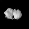 28963MC  7tuwC 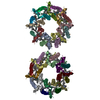 7tv3C 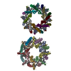 8fb9C M: map data used to model this data C: citing same article ( |
|---|---|
| Similar structure data |
-Links
 PDBj
PDBj
- Assembly
| Deposited unit | 
|
|---|---|
| 1 |
|
-Components
-Light-harvesting protein B800-820 ... , 2 types, 16 molecules QSUWYaceRTVXZbdf
| #1: Protein | Mass: 6088.120 Da / Num. of mol.: 8 / Source method: isolated from a natural source Source: (natural) Magnetospirillum molischianum (magnetotactic)References: UniProt: Q7M119 #2: Protein/peptide | Mass: 5120.873 Da / Num. of mol.: 8 / Source method: isolated from a natural source Source: (natural) Magnetospirillum molischianum (magnetotactic)References: UniProt: Q7M158 |
|---|
-Light-harvesting protein B-800/850 ... , 2 types, 16 molecules gCDGIKMOBEFHJLNP
| #3: Protein | Mass: 5944.980 Da / Num. of mol.: 8 / Source method: isolated from a natural source Source: (natural) Magnetospirillum molischianum (magnetotactic)References: UniProt: P97253 #4: Protein/peptide | Mass: 5120.873 Da / Num. of mol.: 8 / Source method: isolated from a natural source Source: (natural) Magnetospirillum molischianum (magnetotactic)References: UniProt: P95673 |
|---|
-Non-polymers , 2 types, 64 molecules 

| #5: Chemical | ChemComp-BCL /  Mass: 911.504 Da / Num. of mol.: 48 / Source method: obtained synthetically / Formula: C55H74MgN4O6 / Feature type: SUBJECT OF INVESTIGATION Mass: 911.504 Da / Num. of mol.: 48 / Source method: obtained synthetically / Formula: C55H74MgN4O6 / Feature type: SUBJECT OF INVESTIGATION#6: Chemical | ChemComp-LYC /  Mass: 536.873 Da / Num. of mol.: 16 / Source method: obtained synthetically / Formula: C40H56 / Feature type: SUBJECT OF INVESTIGATION Mass: 536.873 Da / Num. of mol.: 16 / Source method: obtained synthetically / Formula: C40H56 / Feature type: SUBJECT OF INVESTIGATION |
|---|
-Details
| Has ligand of interest | Y |
|---|
-Experimental details
-Experiment
| Experiment | Method: ELECTRON MICROSCOPY |
|---|---|
| EM experiment | Aggregation state: PARTICLE / 3D reconstruction method: single particle reconstruction |
- Sample preparation
| Component | Name: LHII-LHIII complex in nanodisk / Type: COMPLEX / Entity ID: #1-#4 / Source: NATURAL |
|---|---|
| Molecular weight | Value: 0.2 MDa / Experimental value: NO |
| Source (natural) | Organism: Magnetospirillum molischianum (magnetotactic) |
| Buffer solution | pH: 7.4 |
| Specimen | Conc.: 1 mg/ml / Embedding applied: NO / Shadowing applied: NO / Staining applied: NO / Vitrification applied: YES |
| Vitrification | Cryogen name: ETHANE |
- Electron microscopy imaging
| Experimental equipment |  Model: Titan Krios / Image courtesy: FEI Company |
|---|---|
| Microscopy | Model: FEI TITAN KRIOS |
| Electron gun | Electron source:  FIELD EMISSION GUN / Accelerating voltage: 300 kV / Illumination mode: FLOOD BEAM FIELD EMISSION GUN / Accelerating voltage: 300 kV / Illumination mode: FLOOD BEAM |
| Electron lens | Mode: BRIGHT FIELD / Nominal defocus max: 2500 nm / Nominal defocus min: 750 nm |
| Image recording | Electron dose: 75 e/Å2 / Film or detector model: GATAN K3 (6k x 4k) |
- Processing
| EM software |
| ||||||||||||||||||||
|---|---|---|---|---|---|---|---|---|---|---|---|---|---|---|---|---|---|---|---|---|---|
| CTF correction | Type: PHASE FLIPPING ONLY | ||||||||||||||||||||
| 3D reconstruction | Resolution: 11.3 Å / Resolution method: FSC 0.143 CUT-OFF / Num. of particles: 11133 / Symmetry type: POINT | ||||||||||||||||||||
| Atomic model building | Protocol: RIGID BODY FIT / Space: REAL |
