 Movie
Movie Controller


+ Open data

- Basic information
| Entry | Database: PDB / ID: 8enu | ||||||
|---|---|---|---|---|---|---|---|
| Title | Structure of the C3bB proconvertase in complex with lufaxin | ||||||
 Components Components |
| ||||||
 Keywords Keywords | IMMUNE SYSTEM / Complement / Alternative pathway / inhibitor / sand fly | ||||||
| Function / homology |  Function and homology information Function and homology informationalternative-complement-pathway C3/C5 convertase / oviduct epithelium development / C5L2 anaphylatoxin chemotactic receptor binding / regulation of triglyceride biosynthetic process / positive regulation of activation of membrane attack complex / complement binding / vertebrate eye-specific patterning / positive regulation of apoptotic cell clearance / complement-mediated synapse pruning / Alternative complement activation ...alternative-complement-pathway C3/C5 convertase / oviduct epithelium development / C5L2 anaphylatoxin chemotactic receptor binding / regulation of triglyceride biosynthetic process / positive regulation of activation of membrane attack complex / complement binding / vertebrate eye-specific patterning / positive regulation of apoptotic cell clearance / complement-mediated synapse pruning / Alternative complement activation / positive regulation of lipid storage / positive regulation of phagocytosis, engulfment / positive regulation of G protein-coupled receptor signaling pathway / complement receptor mediated signaling pathway / Activation of C3 and C5 / positive regulation of type IIa hypersensitivity / positive regulation of glucose transmembrane transport / complement-dependent cytotoxicity / complement activation, alternative pathway / complement activation / endopeptidase inhibitor activity / neuron remodeling / amyloid-beta clearance / positive regulation of vascular endothelial growth factor production / Purinergic signaling in leishmaniasis infection / fatty acid metabolic process / Peptide ligand-binding receptors / complement activation, classical pathway / Regulation of Complement cascade / Post-translational protein phosphorylation / response to bacterium / positive regulation of receptor-mediated endocytosis / positive regulation of angiogenesis / Regulation of Insulin-like Growth Factor (IGF) transport and uptake by Insulin-like Growth Factor Binding Proteins (IGFBPs) / Immunoregulatory interactions between a Lymphoid and a non-Lymphoid cell / azurophil granule lumen / G alpha (i) signalling events / secretory granule lumen / toxin activity / blood microparticle / positive regulation of protein phosphorylation / inflammatory response / immune response / G protein-coupled receptor signaling pathway / endoplasmic reticulum lumen / serine-type endopeptidase activity / signaling receptor binding / Neutrophil degranulation / cell surface / signal transduction / protein-containing complex / proteolysis / extracellular space / extracellular exosome / extracellular region / plasma membrane Similarity search - Function | ||||||
| Biological species |  Lutzomyia longipalpis (insect) Lutzomyia longipalpis (insect) Homo sapiens (human) Homo sapiens (human) | ||||||
| Method | ELECTRON MICROSCOPY / single particle reconstruction / cryo EM / Resolution: 3.22 Å | ||||||
 Authors Authors | Andersen, J.F. / Lei, H. | ||||||
| Funding support |  United States, 1items United States, 1items
| ||||||
 Citation Citation | Journal: Blood / Year: 2023 Title: A bispecific inhibitor of complement and coagulation blocks activation in complementopathy models via a novel mechanism. Authors: John F Andersen / Haotian Lei / Ethan C Strayer / Tapan Kanai / Van Pham / Xiang-Zuo Pan / Patricia Hessab Alvarenga / Gloria F Gerber / Oluwatoyin A Asojo / Ivo M B Francischetti / Robert A ...Authors: John F Andersen / Haotian Lei / Ethan C Strayer / Tapan Kanai / Van Pham / Xiang-Zuo Pan / Patricia Hessab Alvarenga / Gloria F Gerber / Oluwatoyin A Asojo / Ivo M B Francischetti / Robert A Brodsky / Jesus G Valenzuela / José M C Ribeiro / Abstract: Inhibitors of complement and coagulation are present in the saliva of a variety of blood-feeding arthropods that transmit parasitic and viral pathogens. Here, we describe the structure and mechanism ...Inhibitors of complement and coagulation are present in the saliva of a variety of blood-feeding arthropods that transmit parasitic and viral pathogens. Here, we describe the structure and mechanism of action of the sand fly salivary protein lufaxin, which inhibits the formation of the central alternative C3 convertase (C3bBb) and inhibits coagulation factor Xa (fXa). Surface plasmon resonance experiments show that lufaxin stabilizes the binding of serine protease factor B (FB) to C3b but does not detectably bind either C3b or FB alone. The crystal structure of the inhibitor reveals a novel all β-sheet fold containing 2 domains. A structure of the lufaxin-C3bB complex obtained via cryo-electron microscopy (EM) shows that lufaxin binds via its N-terminal domain at an interface containing elements of both C3b and FB. By occupying this spot, the inhibitor locks FB into a closed conformation in which proteolytic activation of FB by FD cannot occur. C3bB-bound lufaxin binds fXa at a separate site in its C-terminal domain. In the cryo-EM structure of a C3bB-lufaxin-fXa complex, the inhibitor binds to both targets simultaneously, and lufaxin inhibits fXa through substrate-like binding of a C-terminal peptide at the active site as well as other interactions in this region. Lufaxin inhibits complement activation in ex vivo models of atypical hemolytic uremic syndrome (aHUS) and paroxysmal nocturnal hemoglobinuria (PNH) as well as thrombin generation in plasma, providing a rationale for the development of a bispecific inhibitor to treat complement-related diseases in which thrombosis is a prominent manifestation. | ||||||
| History |
|
- Structure visualization
| Structure viewer | Molecule: MolmilJmol/JSmol |
|---|
- Downloads & links
-Download
| PDBx/mmCIF format | 8enu.cif.gz | 490.7 KB | Display | PDBx/mmCIF format |
|---|---|---|---|---|
| PDB format | pdb8enu.ent.gz | 384.1 KB | Display | PDB format |
| PDBx/mmJSON format | 8enu.json.gz | Tree view | PDBx/mmJSON format | |
| Others |  Other downloads Other downloads |
-Validation report
| Arichive directory | https://data.pdbj.org/pub/pdb/validation_reports/en/8enuftp://data.pdbj.org/pub/pdb/validation_reports/en/8enu | HTTPS FTP |
|---|
-Related structure data
| Related structure data |  28279MC  8eo2C  8eokC M: map data used to model this data C: citing same article ( |
|---|---|
| Similar structure data |

-Links
 PDBj
PDBj


- Assembly
| Deposited unit | 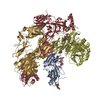
|
|---|---|
| 1 |
|
-Components
-Protein , 4 types, 4 molecules GHDA
| #1: Protein | Mass: 71393.320 Da / Num. of mol.: 1 / Source method: isolated from a natural source / Source: (natural) Homo sapiens (human) / References: UniProt: P01024 |
|---|---|
| #2: Protein | Mass: 104074.148 Da / Num. of mol.: 1 / Source method: isolated from a natural source / Source: (natural) Homo sapiens (human) / References: UniProt: P01024 |
| #3: Protein | Mass: 85510.617 Da / Num. of mol.: 1 / Source method: isolated from a natural source / Source: (natural) Homo sapiens (human)References: UniProt: P00751, alternative-complement-pathway C3/C5 convertase |
| #4: Protein | Mass: 32525.715 Da / Num. of mol.: 1 Source method: isolated from a genetically manipulated source Source: (gene. exp.) Lutzomyia longipalpis (insect) / Cell line (production host): HEK293 / Production host: Homo sapiens (human) / References: UniProt: Q5WPU8 |
-Sugars , 3 types, 7 molecules 
| #5: Polysaccharide | Source method: isolated from a genetically manipulated source #6: Polysaccharide | beta-D-mannopyranose-(1-4)-2-acetamido-2-deoxy-beta-D-glucopyranose-(1-6)-2-acetamido-2-deoxy-beta- ...beta-D-mannopyranose-(1-4)-2-acetamido-2-deoxy-beta-D-glucopyranose-(1-6)-2-acetamido-2-deoxy-beta-D-glucopyranose | Source method: isolated from a genetically manipulated source #7: Sugar | ChemComp-NAG /  Type: D-saccharide, beta linking / Mass: 221.208 Da / Num. of mol.: 4 Type: D-saccharide, beta linking / Mass: 221.208 Da / Num. of mol.: 4Source method: isolated from a genetically manipulated source Formula: C8H15NO6 |
|---|
-Details
| Has ligand of interest | N |
|---|
-Experimental details
-Experiment
| Experiment | Method: ELECTRON MICROSCOPY |
|---|---|
| EM experiment | Aggregation state: PARTICLE / 3D reconstruction method: single particle reconstruction |
- Sample preparation
| Component | Name: The C3 proconvertase from the alternative pathway of complement in complex with lufaxin, a complement inhibitor Type: COMPLEX / Entity ID: #1-#4 / Source: MULTIPLE SOURCES | ||||||||||||||||||||
|---|---|---|---|---|---|---|---|---|---|---|---|---|---|---|---|---|---|---|---|---|---|
| Molecular weight | Value: 0.3023 MDa / Experimental value: YES | ||||||||||||||||||||
| Source (natural) | Organism: Homo sapiens (human) | ||||||||||||||||||||
| Source (recombinant) | Organism: Homo sapiens (human) / Cell: HEK 293 / Plasmid: VR2001 | ||||||||||||||||||||
| Buffer solution | pH: 7.5 | ||||||||||||||||||||
| Buffer component |
| ||||||||||||||||||||
| Specimen | Conc.: 1.5 mg/ml / Embedding applied: NO / Shadowing applied: NO / Staining applied: NO / Vitrification applied: YES / Details: The sample was monodisperse | ||||||||||||||||||||
| Specimen support | Grid material: GOLD / Grid mesh size: 300 divisions/in. / Grid type: C-flat-1.2/1.3 | ||||||||||||||||||||
| Vitrification | Cryogen name: ETHANE |
- Electron microscopy imaging
| Microscopy | Model: TFS GLACIOS |
|---|---|
| Electron gun | Electron source:  FIELD EMISSION GUN / Accelerating voltage: 200 kV / Illumination mode: FLOOD BEAM FIELD EMISSION GUN / Accelerating voltage: 200 kV / Illumination mode: FLOOD BEAM |
| Electron lens | Mode: BRIGHT FIELD / Nominal defocus max: 2200 nm / Nominal defocus min: 300 nm |
| Image recording | Electron dose: 58.31 e/Å2 / Film or detector model: GATAN K3 (6k x 4k) |
- Processing
| EM software |
| |||||||||
|---|---|---|---|---|---|---|---|---|---|---|
| CTF correction | Type: PHASE FLIPPING AND AMPLITUDE CORRECTION | |||||||||
| 3D reconstruction | Resolution: 3.22 Å / Resolution method: FSC 0.143 CUT-OFF / Num. of particles: 80727 / Symmetry type: POINT |
