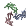
Journal: Biochemistry / Year: 2023 Title: Design of Diverse Asymmetric Pockets in Homo-oligomeric Proteins. Authors: Stacey R Gerben / Andrew J Borst / Derrick R Hicks / Isabelle Moczygemba / David Feldman / Brian Coventry / Wei Yang / Asim K Bera / Marcos Miranda / Alex Kang / Hannah Nguyen / David Baker / Abstract: A challenge for design of protein-small-molecule recognition is that incorporation of cavities with size, shape, and composition suitable for specific recognition can considerably destabilize protein ...A challenge for design of protein-small-molecule recognition is that incorporation of cavities with size, shape, and composition suitable for specific recognition can considerably destabilize protein monomers. This challenge can be overcome through binding pockets formed at homo-oligomeric interfaces between folded monomers. Interfaces surrounding the central homo-oligomer symmetry axes necessarily have the same symmetry and so may not be well suited to binding asymmetric molecules. To enable general recognition of arbitrary asymmetric substrates and small molecules, we developed an approach to designing asymmetric interfaces at off-axis sites on homo-oligomers, analogous to those found in native homo-oligomeric proteins such as glutamine synthetase. We symmetrically dock curved helical repeat proteins such that they form pockets at the asymmetric interface of the oligomer with sizes ranging from several angstroms, appropriate for binding a single ion, to up to more than 20 Å across. Of the 133 proteins tested, 84 had soluble expression in , 47 had correct oligomeric states in solution, 35 had small-angle X-ray scattering (SAXS) data largely consistent with design models, and 8 had negative-stain electron microscopy (nsEM) 2D class averages showing the structures coming together as designed. Both an X-ray crystal structure and a cryogenic electron microscopy (cryoEM) structure are close to the computational design models. The nature of these proteins as homo-oligomers allows them to be readily built into higher-order structures such as nanocages, and the asymmetric pockets of these structures open rich possibilities for small-molecule binder design free from the constraints associated with monomer destabilization.
History
Deposition
Aug 19, 2022
Deposition site: RCSB / Processing site: RCSB
Revision 1.0
Jan 25, 2023
Provider: repository / Type: Initial release
Revision 1.0
Jan 25, 2023
Data content type: EM metadata / Data content type: EM metadata / Provider: repository / Type: Initial release
Revision 1.0
Jan 25, 2023
Data content type: Additional map / Data content type: Additional map / Provider: repository / Type: Initial release
Revision 1.0
Jan 25, 2023
Data content type: FSC / Data content type: FSC / Provider: repository / Type: Initial release
Revision 1.0
Jan 25, 2023
Data content type: Half map / Part number: 1 / Data content type: Half map / Provider: repository / Type: Initial release
Revision 1.0
Jan 25, 2023
Data content type: Half map / Part number: 2 / Data content type: Half map / Provider: repository / Type: Initial release
Revision 1.0
Jan 25, 2023
Data content type: Image / Data content type: Image / Provider: repository / Type: Initial release
Revision 1.0
Jan 25, 2023
Data content type: Primary map / Data content type: Primary map / Provider: repository / Type: Initial release
Revision 1.0
Jan 25, 2023
Data content type: Additional map / Data content type: Additional map / Provider: repository / Type: Initial release
Revision 1.0
Jan 25, 2023
Data content type: FSC / Data content type: FSC / Provider: repository / Type: Initial release
Revision 1.0
Jan 25, 2023
Data content type: Half map / Part number: 1 / Data content type: Half map / Provider: repository / Type: Initial release
Revision 1.0
Jan 25, 2023
Data content type: Half map / Part number: 2 / Data content type: Half map / Provider: repository / Type: Initial release
Revision 1.0
Jan 25, 2023
Data content type: Image / Data content type: Image / Provider: repository / Type: Initial release
Revision 1.0
Jan 25, 2023
Data content type: Primary map / Data content type: Primary map / Provider: repository / Type: Initial release
Revision 1.1
Jun 19, 2024
Group: Data collection / Category: chem_comp_atom / chem_comp_bond
Data content type: EM metadata / Data content type: EM metadata / EM metadata / Group: Data processing / Experimental summary / Data content type: EM metadata / EM metadata / Category: em_admin / em_software / Data content type: EM metadata / EM metadata / Item: _em_admin.last_update / _em_software.name
Evidence: electron microscopy, negative stain EM, cryoEM
Type
Name
Symmetry operation
Number
identity operation
1_555
1
-
Components
#1: Protein
SG135
Mass: 23521.809 Da / Num. of mol.: 4 Source method: isolated from a genetically manipulated source Details: Deleted loop consisting of residues 173-179 due to lack of confident map density Source: (gene. exp.) synthetic construct (others) / Production host: Escherichia coli (E. coli)
Has protein modification
N
-
Experimental details
-
Experiment
Experiment
Method: ELECTRON MICROSCOPY
EM experiment
Aggregation state: PARTICLE / 3D reconstruction method: single particle reconstruction
Resolution: 3.85 Å / Resolution method: FSC 0.143 CUT-OFF / Num. of particles: 855664 Details: Removed large number over over-represented views from initial set of particles. Symmetry type: POINT
Refine LS restraints
Refine-ID
Type
Dev ideal
Number
ELECTRONMICROSCOPY
f_bond_d
0.007
6092
ELECTRONMICROSCOPY
f_angle_d
0.65
8164
ELECTRONMICROSCOPY
f_dihedral_angle_d
12.357
2408
ELECTRONMICROSCOPY
f_chiral_restr
0.03
1004
ELECTRONMICROSCOPY
f_plane_restr
0.004
1044
+
About Yorodumi
-
News
-
Feb 9, 2022. New format data for meta-information of EMDB entries
New format data for meta-information of EMDB entries
Version 3 of the EMDB header file is now the official format.
The previous official version 1.9 will be removed from the archive.
In the structure databanks used in Yorodumi, some data are registered as the other names, "COVID-19 virus" and "2019-nCoV". Here are the details of the virus and the list of structure data.
Jan 31, 2019. EMDB accession codes are about to change! (news from PDBe EMDB page)
EMDB accession codes are about to change! (news from PDBe EMDB page)
The allocation of 4 digits for EMDB accession codes will soon come to an end. Whilst these codes will remain in use, new EMDB accession codes will include an additional digit and will expand incrementally as the available range of codes is exhausted. The current 4-digit format prefixed with “EMD-” (i.e. EMD-XXXX) will advance to a 5-digit format (i.e. EMD-XXXXX), and so on. It is currently estimated that the 4-digit codes will be depleted around Spring 2019, at which point the 5-digit format will come into force.
The EM Navigator/Yorodumi systems omit the EMD- prefix.
Related info.:Q: What is EMD? / ID/Accession-code notation in Yorodumi/EM Navigator
Yorodumi is a browser for structure data from EMDB, PDB, SASBDB, etc.
This page is also the successor to EM Navigator detail page, and also detail information page/front-end page for Omokage search.
The word "yorodu" (or yorozu) is an old Japanese word meaning "ten thousand". "mi" (miru) is to see.
Related info.:EMDB / PDB / SASBDB / Comparison of 3 databanks / Yorodumi Search / Aug 31, 2016. New EM Navigator & Yorodumi / Yorodumi Papers / Jmol/JSmol / Function and homology information / Changes in new EM Navigator and Yorodumi
 Movie
Movie Controller

 Yorodumi
 Open data
 Basic information
 Components
 Keywords
 Authors
 United States, 1items
  Citation
 Structure visualization
 Molmil
 Downloads & links
 Other downloads
 PDBj
PDBj
 Assembly
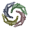

 Sample preparation
 Electron microscopy imaging
 FIELD EMISSION GUN / Accelerating voltage: 300 kV / Illumination mode: FLOOD BEAM
 Processing