+ Open data
- Basic information
| Entry | Database: PDB / ID: 8e2a | |||||||||
|---|---|---|---|---|---|---|---|---|---|---|
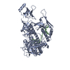
| Title | Human Dis3L2 in complex with hairpin D-U7 | |||||||||
 Components Components |
| |||||||||
 Keywords Keywords | RNA BINDING PROTEIN/RNA / 3'-5' exonuclease / ds-RNA bound exonuclease / human exonuclease / RNA BINDING PROTEIN-RNA complex | |||||||||
| Function / homology |  Function and homology information Function and homology informationpolyuridylation-dependent mRNA catabolic process / miRNA catabolic process / mitotic sister chromatid separation / Z-decay: degradation of maternal mRNAs by zygotically expressed factors / poly(U) RNA binding / stem cell population maintenance / mRNA catabolic process / nuclear-transcribed mRNA catabolic process / RNA nuclease activity / P-body ...polyuridylation-dependent mRNA catabolic process / miRNA catabolic process / mitotic sister chromatid separation / Z-decay: degradation of maternal mRNAs by zygotically expressed factors / poly(U) RNA binding / stem cell population maintenance / mRNA catabolic process / nuclear-transcribed mRNA catabolic process / RNA nuclease activity / P-body / mitotic cell cycle / Hydrolases; Acting on ester bonds; Exoribonucleases producing 5'-phosphomonoesters / 3'-5'-RNA exonuclease activity / negative regulation of cell population proliferation / cell division / magnesium ion binding / cytoplasm Similarity search - Function | |||||||||
| Biological species |  Homo sapiens (human) Homo sapiens (human)synthetic construct (others) | |||||||||
| Method | ELECTRON MICROSCOPY / single particle reconstruction / cryo EM / Resolution: 2.8 Å | |||||||||
 Authors Authors | Meze, K. / Thomas, D.R. / Joshua-Tor, L. | |||||||||
| Funding support |  United States, 2items United States, 2items
| |||||||||
 Citation Citation |  Journal: Nat Struct Mol Biol / Year: 2023 Journal: Nat Struct Mol Biol / Year: 2023Title: A shape-shifting nuclease unravels structured RNA. Authors: Katarina Meze / Armend Axhemi / Dennis R Thomas / Ahmet Doymaz / Leemor Joshua-Tor /  Abstract: RNA turnover pathways ensure appropriate gene expression levels by eliminating unwanted transcripts. Dis3-like 2 (Dis3L2) is a 3'-5' exoribonuclease that plays a critical role in human development. ...RNA turnover pathways ensure appropriate gene expression levels by eliminating unwanted transcripts. Dis3-like 2 (Dis3L2) is a 3'-5' exoribonuclease that plays a critical role in human development. Dis3L2 independently degrades structured substrates, including coding and noncoding 3' uridylated RNAs. While the basis for Dis3L2's substrate recognition has been well characterized, the mechanism of structured RNA degradation by this family of enzymes is unknown. We characterized the discrete steps of the degradation cycle by determining cryogenic electron microscopy structures representing snapshots along the RNA turnover pathway and measuring kinetic parameters for RNA processing. We discovered a dramatic conformational change that is triggered by double-stranded RNA (dsRNA), repositioning two cold shock domains by 70 Å. This movement exposes a trihelix linker region, which acts as a wedge to separate the two RNA strands. Furthermore, we show that the trihelix linker is critical for dsRNA, but not single-stranded RNA, degradation. These findings reveal the conformational plasticity of Dis3L2 and detail a mechanism of structured RNA degradation. | |||||||||
| History |
|
- Structure visualization
| Structure viewer | Molecule:  Molmil Molmil Jmol/JSmol Jmol/JSmol |
|---|
- Downloads & links
- Download
| PDBx/mmCIF format |  8e2a.cif.gz 8e2a.cif.gz | 145.4 KB | Display |  PDBx/mmCIF format PDBx/mmCIF format |
|---|---|---|---|---|
| PDB format |  pdb8e2a.ent.gz pdb8e2a.ent.gz | 107.4 KB | Display |  PDB format PDB format |
| PDBx/mmJSON format |  8e2a.json.gz 8e2a.json.gz | Tree view |  PDBx/mmJSON format PDBx/mmJSON format | |
| Others |  Other downloads Other downloads |
-Validation report
| Arichive directory |  https://data.pdbj.org/pub/pdb/validation_reports/e2/8e2a https://data.pdbj.org/pub/pdb/validation_reports/e2/8e2a ftp://data.pdbj.org/pub/pdb/validation_reports/e2/8e2a ftp://data.pdbj.org/pub/pdb/validation_reports/e2/8e2a | HTTPS FTP |
|---|
-Related structure data
| Related structure data |  27830MC  8e27C  8e28C  8e29C M: map data used to model this data C: citing same article ( |
|---|---|
| Similar structure data | Similarity search - Function & homology  F&H Search F&H Search |
- Links
Links
- Assembly
| Deposited unit | 
|
|---|---|
| 1 |
|
- Components
| #1: Protein | Mass: 99414.195 Da / Num. of mol.: 1 Source method: isolated from a genetically manipulated source Source: (gene. exp.)  Homo sapiens (human) / Gene: DIS3L2, FAM6A / Production host: Homo sapiens (human) / Gene: DIS3L2, FAM6A / Production host:  References: UniProt: Q8IYB7, Hydrolases; Acting on ester bonds; Exoribonucleases producing 5'-phosphomonoesters |
|---|---|
| #2: RNA chain | Mass: 7248.232 Da / Num. of mol.: 1 / Source method: obtained synthetically Details: The full sequence of the hairpin in the sample is: CGGCGUUUCGACGCCGUUUUUUU. Some of the hairpin, including the tetraloop, was not resolved in the structure. Source: (synth.) synthetic construct (others) |
-Experimental details
-Experiment
| Experiment | Method: ELECTRON MICROSCOPY |
|---|---|
| EM experiment | Aggregation state: PARTICLE / 3D reconstruction method: single particle reconstruction |
- Sample preparation
| Component | Name: Wild-type HsDis3L2 in complex with hairpinD-U7 / Type: COMPLEX / Entity ID: all / Source: MULTIPLE SOURCES | ||||||||||||||||||||
|---|---|---|---|---|---|---|---|---|---|---|---|---|---|---|---|---|---|---|---|---|---|
| Molecular weight | Value: 0.1 MDa / Experimental value: NO | ||||||||||||||||||||
| Source (natural) |
| ||||||||||||||||||||
| Source (recombinant) | Organism:  | ||||||||||||||||||||
| Buffer solution | pH: 7.5 | ||||||||||||||||||||
| Buffer component |
| ||||||||||||||||||||
| Specimen | Conc.: 0.3 mg/ml / Embedding applied: NO / Shadowing applied: NO / Staining applied: NO / Vitrification applied: YES | ||||||||||||||||||||
| Specimen support | Grid material: GOLD / Grid type: UltrAuFoil R1.2/1.3 | ||||||||||||||||||||
| Vitrification | Instrument: FEI VITROBOT MARK IV / Cryogen name: ETHANE / Humidity: 90 % / Chamber temperature: 295 K |
- Electron microscopy imaging
| Experimental equipment |  Model: Titan Krios / Image courtesy: FEI Company |
|---|---|
| Microscopy | Model: FEI TITAN KRIOS |
| Electron gun | Electron source:  FIELD EMISSION GUN / Accelerating voltage: 300 kV / Illumination mode: FLOOD BEAM FIELD EMISSION GUN / Accelerating voltage: 300 kV / Illumination mode: FLOOD BEAM |
| Electron lens | Mode: BRIGHT FIELD / Nominal magnification: 130000 X / Nominal defocus max: 2600 nm / Nominal defocus min: 700 nm / Calibrated defocus min: 500 nm / Calibrated defocus max: 2800 nm / Cs: 2.7 mm / C2 aperture diameter: 70 µm / Alignment procedure: ZEMLIN TABLEAU |
| Specimen holder | Cryogen: NITROGEN / Specimen holder model: FEI TITAN KRIOS AUTOGRID HOLDER / Temperature (max): 80 K / Temperature (min): 80 K |
| Image recording | Electron dose: 62.4 e/Å2 / Detector mode: COUNTING / Film or detector model: GATAN K3 BIOQUANTUM (6k x 4k) / Num. of grids imaged: 1 / Num. of real images: 4662 |
| Image scans | Sampling size: 5 µm / Width: 5760 / Height: 4092 / Movie frames/image: 30 / Used frames/image: 1-30 |
- Processing
| Software | Name: PHENIX / Version: 1.18_3855: / Classification: refinement | ||||||||||||||||||||||||||||||||||||||||
|---|---|---|---|---|---|---|---|---|---|---|---|---|---|---|---|---|---|---|---|---|---|---|---|---|---|---|---|---|---|---|---|---|---|---|---|---|---|---|---|---|---|
| EM software |
| ||||||||||||||||||||||||||||||||||||||||
| CTF correction | Type: PHASE FLIPPING ONLY | ||||||||||||||||||||||||||||||||||||||||
| Particle selection | Num. of particles selected: 1836786 | ||||||||||||||||||||||||||||||||||||||||
| Symmetry | Point symmetry: C1 (asymmetric) | ||||||||||||||||||||||||||||||||||||||||
| 3D reconstruction | Resolution: 2.8 Å / Resolution method: FSC 0.143 CUT-OFF / Num. of particles: 539985 / Symmetry type: POINT | ||||||||||||||||||||||||||||||||||||||||
| Atomic model building | Protocol: FLEXIBLE FIT / Space: REAL Details: The model of Homo Sapiens Dis3L2 was used as a starting reference for the protein and the nucleic acid was used from PDB 4PMW. The CSDs were repositioned to fit into the repositioned density ...Details: The model of Homo Sapiens Dis3L2 was used as a starting reference for the protein and the nucleic acid was used from PDB 4PMW. The CSDs were repositioned to fit into the repositioned density via rigid body fit and the linker was rebuilt manually. Base pairing restraints were used for the base of the RNA hairpin for which density was present. All manual building was done in Coot. The model was further refined using PHENIX. | ||||||||||||||||||||||||||||||||||||||||
| Atomic model building | PDB-ID: 4PMW Pdb chain-ID: C / Accession code: 4PMW / Source name: PDB / Type: experimental model |
 Movie
Movie Controller


 PDBj
PDBj


