ムービー
ムービー コントローラー


+ データを開く

- 基本情報
基本情報
| 登録情報 | データベース: PDB / ID: 8dmx | ||||||
|---|---|---|---|---|---|---|---|
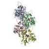
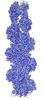
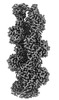
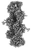
| タイトル | Cryo-EM structure of skeletal muscle alpha-actin | ||||||
 要素 要素 | Actin, alpha skeletal muscle | ||||||
 キーワード キーワード | STRUCTURAL PROTEIN / cytoskeleton | ||||||
| 機能・相同性 |  機能・相同性情報 機能・相同性情報cytoskeletal motor activator activity / tropomyosin binding / mesenchyme migration / troponin I binding / myosin heavy chain binding / filamentous actin / actin filament bundle / skeletal muscle thin filament assembly / striated muscle thin filament / actin filament bundle assembly ...cytoskeletal motor activator activity / tropomyosin binding / mesenchyme migration / troponin I binding / myosin heavy chain binding / filamentous actin / actin filament bundle / skeletal muscle thin filament assembly / striated muscle thin filament / actin filament bundle assembly / skeletal muscle myofibril / actin monomer binding / skeletal muscle fiber development / stress fiber / titin binding / actin filament polymerization / filopodium / actin filament / 加水分解酵素; 酸無水物に作用; 酸無水物に作用・細胞または細胞小器官の運動に関与 / calcium-dependent protein binding / lamellipodium / cell body / hydrolase activity / protein domain specific binding / calcium ion binding / positive regulation of gene expression / magnesium ion binding / ATP binding / identical protein binding / cytoplasm 類似検索 - 分子機能 | ||||||
| 生物種 |  | ||||||
| 手法 | 電子顕微鏡法 / らせん対称体再構成法 / クライオ電子顕微鏡法 / 解像度: 3.37 Å | ||||||
 データ登録者 データ登録者 | Arora, A.S. / Huang, H.L. / Heissler, S.M. / Chinthalapudi, K. | ||||||
| 資金援助 |  米国, 1件 米国, 1件
| ||||||
 引用 引用 | ジャーナル: Elife / 年: 2023 タイトル: Structural insights into actin isoforms. 著者: Amandeep S Arora / Hsiang-Ling Huang / Ramanpreet Singh / Yoshie Narui / Andrejus Suchenko / Tomoyuki Hatano / Sarah M Heissler / Mohan K Balasubramanian / Krishna Chinthalapudi /  要旨: Actin isoforms organize into distinct networks that are essential for the normal function of eukaryotic cells. Despite a high level of sequence and structure conservation, subtle differences in their ...Actin isoforms organize into distinct networks that are essential for the normal function of eukaryotic cells. Despite a high level of sequence and structure conservation, subtle differences in their design principles determine the interaction with myosin motors and actin-binding proteins. Therefore, identifying how the structure of actin isoforms relates to function is important for our understanding of normal cytoskeletal physiology. Here, we report the high-resolution structures of filamentous skeletal muscle α-actin (3.37 Å), cardiac muscle α-actin (3.07 Å), ß-actin (2.99 Å), and γ-actin (3.38 Å) in the Mg·ADP state with their native post-translational modifications. The structures revealed isoform-specific conformations of the N-terminus that shift closer to the filament surface upon myosin binding, thereby establishing isoform-specific interfaces. Collectively, the structures of single-isotype, post-translationally modified bare skeletal muscle α-actin, cardiac muscle α-actin, ß-actin, and γ-actin reveal general principles, similarities, and differences between isoforms. They complement the repertoire of known actin structures and allow for a comprehensive understanding of in vitro and in vivo functions of actin isoforms. | ||||||
| 履歴 |
|
- 構造の表示
構造の表示
| 構造ビューア | 分子: MolmilJmol/JSmol |
|---|
- ダウンロードとリンク
-ダウンロード
| PDBx/mmCIF形式 | 8dmx.cif.gz | 287.4 KB | 表示 | PDBx/mmCIF形式 |
|---|---|---|---|---|
| PDB形式 | pdb8dmx.ent.gz | 236.9 KB | 表示 | PDB形式 |
| PDBx/mmJSON形式 | 8dmx.json.gz | ツリー表示 | PDBx/mmJSON形式 | |
| その他 |  その他のダウンロード その他のダウンロード |
-検証レポート
| 文書・要旨 | 8dmx_validation.pdf.gz | 1.8 MB | 表示 | wwPDB検証レポート |
|---|---|---|---|---|
| 文書・詳細版 | 8dmx_full_validation.pdf.gz | 1.8 MB | 表示 | |
| XML形式データ | 8dmx_validation.xml.gz | 58.3 KB | 表示 | |
| CIF形式データ | 8dmx_validation.cif.gz | 83.7 KB | 表示 | |
| アーカイブディレクトリ | https://data.pdbj.org/pub/pdb/validation_reports/dm/8dmxftp://data.pdbj.org/pub/pdb/validation_reports/dm/8dmx | HTTPS FTP |
-関連構造データ
-リンク
 PDBj
PDBj


- 集合体
集合体
| 登録構造単位 | 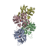
|
|---|---|
| 1 |
|
-要素
| #1: タンパク質 | 分子量: 41875.633 Da / 分子数: 4 / 由来タイプ: 天然 / 由来: (天然) #2: 化合物 | ChemComp-MG /   分子量: 24.305 Da / 分子数: 4 / 由来タイプ: 合成 / 式: Mg 分子量: 24.305 Da / 分子数: 4 / 由来タイプ: 合成 / 式: Mg#3: 化合物 | ChemComp-ADP /   分子量: 427.201 Da / 分子数: 4 / 由来タイプ: 合成 / 式: C10H15N5O10P2 / コメント: ADP, エネルギー貯蔵分子*YM 分子量: 427.201 Da / 分子数: 4 / 由来タイプ: 合成 / 式: C10H15N5O10P2 / コメント: ADP, エネルギー貯蔵分子*YM研究の焦点であるリガンドがあるか | N | |
|---|
-実験情報
-実験
| 実験 | 手法: 電子顕微鏡法 |
|---|---|
| EM実験 | 試料の集合状態: FILAMENT / 3次元再構成法: らせん対称体再構成法 |
- 試料調製
試料調製
| 構成要素 | 名称: actin / タイプ: ORGANELLE OR CELLULAR COMPONENT / Entity ID: #1 / 由来: NATURAL |
|---|---|
| 分子量 | 実験値: NO |
| 由来(天然) | 生物種: |
| 緩衝液 | pH: 7 |
| 試料 | 包埋: NO / シャドウイング: NO / 染色: NO / 凍結: YES |
| 試料支持 | グリッドの材料: GOLD / グリッドのタイプ: C-flat |
| 急速凍結 | 装置: LEICA EM GP / 凍結剤: ETHANE / 湿度: 95 % / 凍結前の試料温度: 298 K |
- 電子顕微鏡撮影
| 実験機器 |  モデル: Titan Krios / 画像提供: FEI Company |
|---|---|
| 顕微鏡 | モデル: FEI TITAN KRIOS |
| 電子銃 | 電子線源:  FIELD EMISSION GUN / 加速電圧: 300 kV / 照射モード: OTHER FIELD EMISSION GUN / 加速電圧: 300 kV / 照射モード: OTHER |
| 電子レンズ | モード: BRIGHT FIELD / 倍率(公称値): 81000 X / 最大 デフォーカス(公称値): 1500 nm / 最小 デフォーカス(公称値): 200 nm / アライメント法: ZEMLIN TABLEAU |
| 試料ホルダ | 凍結剤: NITROGEN 試料ホルダーモデル: FEI TITAN KRIOS AUTOGRID HOLDER |
| 撮影 | 電子線照射量: 65 e/Å2 フィルム・検出器のモデル: GATAN K3 BIOQUANTUM (6k x 4k) 撮影したグリッド数: 1 / 実像数: 2046 |
| 電子光学装置 | エネルギーフィルター名称: GIF Bioquantum / エネルギーフィルタースリット幅: 20 eV / 球面収差補正装置: Cs-corrected microscope |
- 解析
解析
| ソフトウェア | 名称: PHENIX / バージョン: 1.20rc3_4406: / 分類: 精密化 | ||||||||||||||||||||||||||||||
|---|---|---|---|---|---|---|---|---|---|---|---|---|---|---|---|---|---|---|---|---|---|---|---|---|---|---|---|---|---|---|---|
| EMソフトウェア |
| ||||||||||||||||||||||||||||||
| CTF補正 | タイプ: NONE | ||||||||||||||||||||||||||||||
| らせん対称 | 回転角度/サブユニット: -166 ° / 軸方向距離/サブユニット: 28 Å / らせん対称軸の対称性: C1 | ||||||||||||||||||||||||||||||
| 粒子像の選択 | 詳細: Single Particle | ||||||||||||||||||||||||||||||
| 3次元再構成 | 解像度: 3.37 Å / 解像度の算出法: FSC 0.143 CUT-OFF / 粒子像の数: 261195 / 対称性のタイプ: HELICAL | ||||||||||||||||||||||||||||||
| 原子モデル構築 | B value: 90 / プロトコル: FLEXIBLE FIT / 空間: REAL | ||||||||||||||||||||||||||||||
| 原子モデル構築 | PDB-ID: 6DJO PDB chain-ID: A | ||||||||||||||||||||||||||||||
| 拘束条件 |
|