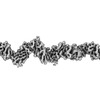
ジャーナル: PLoS Pathog / 年: 2023 タイトル: Architecture of the biofilm-associated archaic Chaperone-Usher pilus CupE from Pseudomonas aeruginosa. 著者: Jan Böhning / Adrian W Dobbelstein / Nina Sulkowski / Kira Eilers / Andriko von Kügelgen / Abul K Tarafder / Sew-Yeu Peak-Chew / Mark Skehel / Vikram Alva / Alain Filloux / Tanmay A M Bharat / 要旨: Chaperone-Usher Pathway (CUP) pili are major adhesins in Gram-negative bacteria, mediating bacterial adherence to biotic and abiotic surfaces. While classical CUP pili have been extensively ...Chaperone-Usher Pathway (CUP) pili are major adhesins in Gram-negative bacteria, mediating bacterial adherence to biotic and abiotic surfaces. While classical CUP pili have been extensively characterized, little is known about so-called archaic CUP pili, which are phylogenetically widespread and promote biofilm formation by several human pathogens. In this study, we present the electron cryomicroscopy structure of the archaic CupE pilus from the opportunistic human pathogen Pseudomonas aeruginosa. We show that CupE1 subunits within the pilus are arranged in a zigzag architecture, containing an N-terminal donor β-strand extending from each subunit into the next, where it is anchored by hydrophobic interactions, with comparatively weaker interactions at the rest of the inter-subunit interface. Imaging CupE pili on the surface of P. aeruginosa cells using electron cryotomography shows that CupE pili adopt variable curvatures in response to their environment, which might facilitate their role in promoting cellular attachment. Finally, bioinformatic analysis shows the widespread abundance of cupE genes in isolates of P. aeruginosa and the co-occurrence of cupE with other cup clusters, suggesting interdependence of cup pili in regulating bacterial adherence within biofilms. Taken together, our study provides insights into the architecture of archaic CUP pili, providing a structural basis for understanding their role in promoting cellular adhesion and biofilm formation in P. aeruginosa.
A: SCPU domain-containing protein B: SCPU domain-containing protein C: SCPU domain-containing protein D: SCPU domain-containing protein E: SCPU domain-containing protein
 ムービー
ムービー コントローラー


 データを開く
 基本情報
基本情報 要素
要素 キーワード
キーワード 機能・相同性情報
 Pseudomonas aeruginosa PAO1 (緑膿菌)
 データ登録者
 英国,
英国,  米国, 7件
  引用
引用
 構造の表示
構造の表示 ダウンロードとリンク
 その他のダウンロード
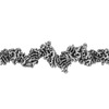
 PDBj
PDBj 集合体
集合体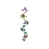
 試料調製
試料調製 Pseudomonas aeruginosa (緑膿菌) / 株: PAO1 mutant / 細胞内の位置: Cell surface
 電子顕微鏡撮影
 FIELD EMISSION GUN / 加速電圧: 300 kV / 照射モード: FLOOD BEAM
 解析
解析