 Movie
Movie Controller

[English] 日本語
 Yorodumi
- PDB-8ca7: Omadacycline and spectinomycin bound to the 30S ribosomal subunit head -
+ Open data

- Basic information
| Entry | Database: PDB / ID: 8ca7 | ||||||||||||
|---|---|---|---|---|---|---|---|---|---|---|---|---|---|
| Title | Omadacycline and spectinomycin bound to the 30S ribosomal subunit head | ||||||||||||
 Components Components |
| ||||||||||||
 Keywords Keywords | RIBOSOME / antibiotics / spectinomycin / omadacycline | ||||||||||||
| Function / homology |  Function and homology information Function and homology informationtranscription antitermination factor activity, RNA binding / regulation of DNA-templated transcription elongation / transcription elongation factor complex / transcription antitermination / maintenance of translational fidelity / ribosomal small subunit assembly / ribosome biogenesis / small ribosomal subunit / cytosolic small ribosomal subunit / cytoplasmic translation ...transcription antitermination factor activity, RNA binding / regulation of DNA-templated transcription elongation / transcription elongation factor complex / transcription antitermination / maintenance of translational fidelity / ribosomal small subunit assembly / ribosome biogenesis / small ribosomal subunit / cytosolic small ribosomal subunit / cytoplasmic translation / tRNA binding / negative regulation of translation / rRNA binding / structural constituent of ribosome / ribosome / translation / response to antibiotic / mRNA binding / RNA binding / membrane / cytosol / cytoplasm Similarity search - Function | ||||||||||||
| Biological species |  | ||||||||||||
| Method | ELECTRON MICROSCOPY / single particle reconstruction / cryo EM / Resolution: 2.06 Å | ||||||||||||
 Authors Authors | Paternoga, H. / Crowe-McAuliffe, C. / Wilson, D.N. | ||||||||||||
| Funding support | European Union, 3items
| ||||||||||||
 Citation Citation | Journal: Nat Struct Mol Biol / Year: 2023 Title: Structural conservation of antibiotic interaction with ribosomes. Authors: Helge Paternoga / Caillan Crowe-McAuliffe / Lars V Bock / Timm O Koller / Martino Morici / Bertrand Beckert / Alexander G Myasnikov / Helmut Grubmüller / Jiří Nováček / Daniel N Wilson /    Abstract: The ribosome is a major target for clinically used antibiotics, but multidrug resistant pathogenic bacteria are making our current arsenal of antimicrobials obsolete. Here we present cryo-electron- ...The ribosome is a major target for clinically used antibiotics, but multidrug resistant pathogenic bacteria are making our current arsenal of antimicrobials obsolete. Here we present cryo-electron-microscopy structures of 17 distinct compounds from six different antibiotic classes bound to the bacterial ribosome at resolutions ranging from 1.6 to 2.2 Å. The improved resolution enables a precise description of antibiotic-ribosome interactions, encompassing solvent networks that mediate multiple additional interactions between the drugs and their target. Our results reveal a high structural conservation in the binding mode between antibiotics with the same scaffold, including ordered water molecules. Water molecules are visualized within the antibiotic binding sites that are preordered, become ordered in the presence of the drug and that are physically displaced on drug binding. Insight into RNA-ligand interactions will facilitate development of new antimicrobial agents, as well as other RNA-targeting therapies. | ||||||||||||
| History |
|
- Structure visualization
| Structure viewer | Molecule: MolmilJmol/JSmol |
|---|
- Downloads & links
-Download
| PDBx/mmCIF format | 8ca7.cif.gz | 510.1 KB | Display | PDBx/mmCIF format |
|---|---|---|---|---|
| PDB format | pdb8ca7.ent.gz | 372.6 KB | Display | PDB format |
| PDBx/mmJSON format | 8ca7.json.gz | Tree view | PDBx/mmJSON format | |
| Others |  Other downloads Other downloads |
-Validation report
| Arichive directory | https://data.pdbj.org/pub/pdb/validation_reports/ca/8ca7ftp://data.pdbj.org/pub/pdb/validation_reports/ca/8ca7 | HTTPS FTP |
|---|
-Related structure data
| Related structure data |  16520MC  8caiC  8camC  8cazC  8cepC  8ceuC  8cf1C  8cf8C  8cgdC  8cgiC  8cgjC  8cgkC  8cgrC 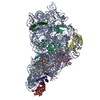 8cguC  8cgvC M: map data used to model this data C: citing same article ( |
|---|---|
| Similar structure data |

-Links
 PDBj
PDBj


- Assembly
| Deposited unit | 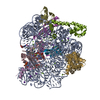
|
|---|---|
| 1 |
|
-Components
-Small ribosomal subunit protein ... , 7 types, 7 molecules CEIJMNS
| #2: Protein | Mass: 26031.316 Da / Num. of mol.: 1 / Source method: isolated from a natural source / Source: (natural) |
|---|---|
| #3: Protein | Mass: 17629.398 Da / Num. of mol.: 1 / Source method: isolated from a natural source / Source: (natural) |
| #5: Protein | Mass: 14886.270 Da / Num. of mol.: 1 / Source method: isolated from a natural source / Source: (natural) |
| #6: Protein | Mass: 11755.597 Da / Num. of mol.: 1 / Source method: isolated from a natural source / Source: (natural) |
| #7: Protein | Mass: 13128.467 Da / Num. of mol.: 1 / Source method: isolated from a natural source / Source: (natural) |
| #8: Protein | Mass: 11606.560 Da / Num. of mol.: 1 / Source method: isolated from a natural source / Source: (natural) |
| #9: Protein | Mass: 10455.355 Da / Num. of mol.: 1 / Source method: isolated from a natural source / Source: (natural) |
-RNA chain / Protein , 2 types, 2 molecules AG
| #1: RNA chain | Mass: 499124.750 Da / Num. of mol.: 1 / Source method: isolated from a natural source / Source: (natural) |
|---|---|
| #4: Protein | Mass: 20055.156 Da / Num. of mol.: 1 / Source method: isolated from a natural source / Source: (natural) |
-Non-polymers , 5 types, 585 molecules 


| #10: Chemical | ChemComp-K /  Mass: 39.098 Da / Num. of mol.: 14 / Source method: obtained synthetically / Formula: K Mass: 39.098 Da / Num. of mol.: 14 / Source method: obtained synthetically / Formula: K#11: Chemical | ChemComp-U3B / |  Mass: 556.650 Da / Num. of mol.: 1 / Source method: obtained synthetically / Formula: C29H40N4O7 / Feature type: SUBJECT OF INVESTIGATION / Comment: medication, antibiotic*YM Mass: 556.650 Da / Num. of mol.: 1 / Source method: obtained synthetically / Formula: C29H40N4O7 / Feature type: SUBJECT OF INVESTIGATION / Comment: medication, antibiotic*YM#12: Chemical | ChemComp-SCM / |  Mass: 332.350 Da / Num. of mol.: 1 / Source method: obtained synthetically / Formula: C14H24N2O7 / Feature type: SUBJECT OF INVESTIGATION Mass: 332.350 Da / Num. of mol.: 1 / Source method: obtained synthetically / Formula: C14H24N2O7 / Feature type: SUBJECT OF INVESTIGATION#13: Chemical | ChemComp-MG /  Mass: 24.305 Da / Num. of mol.: 26 / Source method: obtained synthetically / Formula: Mg Mass: 24.305 Da / Num. of mol.: 26 / Source method: obtained synthetically / Formula: Mg#14: Water | ChemComp-HOH / | Mass: 18.015 Da / Num. of mol.: 543 / Source method: isolated from a natural source / Formula: H2O |
|---|
-Details
| Has ligand of interest | Y |
|---|
-Experimental details
-Experiment
| Experiment | Method: ELECTRON MICROSCOPY |
|---|---|
| EM experiment | Aggregation state: PARTICLE / 3D reconstruction method: single particle reconstruction |
- Sample preparation
| Component | Name: Ribosomal small subunit head / Type: RIBOSOME / Entity ID: #1-#9 / Source: NATURAL | |||||||||||||||||||||||||||||||||||
|---|---|---|---|---|---|---|---|---|---|---|---|---|---|---|---|---|---|---|---|---|---|---|---|---|---|---|---|---|---|---|---|---|---|---|---|---|
| Molecular weight | Experimental value: NO | |||||||||||||||||||||||||||||||||||
| Source (natural) | Organism: | |||||||||||||||||||||||||||||||||||
| Buffer solution | pH: 7.5 | |||||||||||||||||||||||||||||||||||
| Buffer component |
| |||||||||||||||||||||||||||||||||||
| Specimen | Embedding applied: NO / Shadowing applied: NO / Staining applied: NO / Vitrification applied: YES | |||||||||||||||||||||||||||||||||||
| Specimen support | Grid material: GOLD / Grid mesh size: 300 divisions/in. / Grid type: Quantifoil R1.2/1.3 | |||||||||||||||||||||||||||||||||||
| Vitrification | Cryogen name: ETHANE / Humidity: 100 % / Chamber temperature: 277.15 K |
- Electron microscopy imaging
| Experimental equipment |  Model: Titan Krios / Image courtesy: FEI Company |
|---|---|
| Microscopy | Model: FEI TITAN KRIOS |
| Electron gun | Electron source:  FIELD EMISSION GUN / Accelerating voltage: 300 kV / Illumination mode: FLOOD BEAM FIELD EMISSION GUN / Accelerating voltage: 300 kV / Illumination mode: FLOOD BEAM |
| Electron lens | Mode: BRIGHT FIELD / Nominal defocus max: 1600 nm / Nominal defocus min: 400 nm |
| Image recording | Average exposure time: 4 sec. / Electron dose: 44 e/Å2 / Detector mode: COUNTING / Film or detector model: GATAN K2 SUMMIT (4k x 4k) / Num. of real images: 24195 |
- Processing
| EM software | Name: REFMAC / Version: 5.8.0415 / Category: model refinement | ||||||||||||||||||||||||||||||||||||||||||||||||||||||||||||||||||||||||||||||||||||||||||||||||||||||||||
|---|---|---|---|---|---|---|---|---|---|---|---|---|---|---|---|---|---|---|---|---|---|---|---|---|---|---|---|---|---|---|---|---|---|---|---|---|---|---|---|---|---|---|---|---|---|---|---|---|---|---|---|---|---|---|---|---|---|---|---|---|---|---|---|---|---|---|---|---|---|---|---|---|---|---|---|---|---|---|---|---|---|---|---|---|---|---|---|---|---|---|---|---|---|---|---|---|---|---|---|---|---|---|---|---|---|---|---|
| CTF correction | Type: PHASE FLIPPING AND AMPLITUDE CORRECTION | ||||||||||||||||||||||||||||||||||||||||||||||||||||||||||||||||||||||||||||||||||||||||||||||||||||||||||
| 3D reconstruction | Resolution: 2.06 Å / Resolution method: FSC 0.143 CUT-OFF / Num. of particles: 514855 / Symmetry type: POINT | ||||||||||||||||||||||||||||||||||||||||||||||||||||||||||||||||||||||||||||||||||||||||||||||||||||||||||
| Atomic model building | PDB-ID: 7k00 Accession code: 7k00 / Source name: PDB / Type: experimental model | ||||||||||||||||||||||||||||||||||||||||||||||||||||||||||||||||||||||||||||||||||||||||||||||||||||||||||
| Refinement | Resolution: 2.06→134.11 Å / Cor.coef. Fo:Fc: 0.917 / SU B: 4.53 / SU ML: 0.116 / ESU R: 0.134 Stereochemistry target values: MAXIMUM LIKELIHOOD WITH PHASES Details: HYDROGENS HAVE BEEN USED IF PRESENT IN THE INPUT
| ||||||||||||||||||||||||||||||||||||||||||||||||||||||||||||||||||||||||||||||||||||||||||||||||||||||||||
| Solvent computation | Solvent model: PARAMETERS FOR MASK CACLULATION | ||||||||||||||||||||||||||||||||||||||||||||||||||||||||||||||||||||||||||||||||||||||||||||||||||||||||||
| Displacement parameters | Biso mean: 58.887 Å2 | ||||||||||||||||||||||||||||||||||||||||||||||||||||||||||||||||||||||||||||||||||||||||||||||||||||||||||
| Refinement step | Cycle: 1 / Total: 18093 | ||||||||||||||||||||||||||||||||||||||||||||||||||||||||||||||||||||||||||||||||||||||||||||||||||||||||||
| Refine LS restraints |
|
